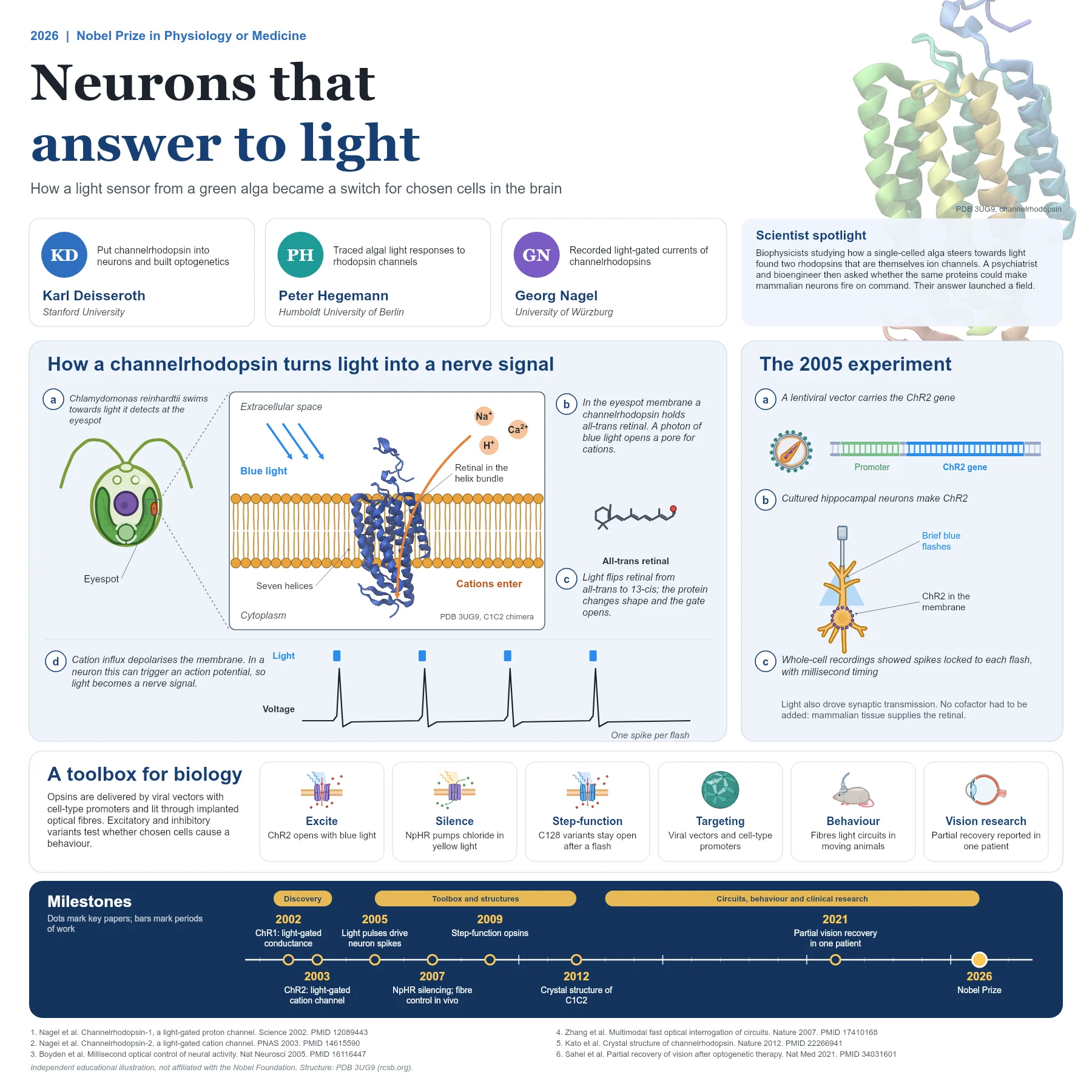
The question: can we control one kind of cell at a time?
The brain contains many intermingled types of neurons, and for decades researchers could record from them far more easily than they could control them. Electrical stimulation excites every cell near the electrode, and drugs act over seconds to minutes. To find out which cells cause a behaviour, neuroscientists needed a switch that was both fast and selective.
The 2026 Nobel Prize in Physiology or Medicine recognises the discovery of light-gated ion channels and their use to build that switch, a method now called optogenetics.
The discovery story: an alga that swims towards light
The single-celled green alga Chlamydomonas reinhardtii swims towards or away from light, guided by an orange eyespot. Biophysicists suspected that rhodopsin-like proteins with a retinal chromophore sensed the light, but how that sensing produced the fast electrical currents seen in the cell was unclear.
In 2002 Georg Nagel, Peter Hegemann and colleagues reported a cDNA from the alga encoding a protein they named channelopsin-1. When they expressed it in frog oocytes with all-trans retinal, light produced a conductance that behaved like a proton channel. The protein, channelrhodopsin-1, was both the light sensor and the channel.
A year later the same team showed that a second protein, channelrhodopsin-2 (ChR2), is a directly light-gated cation channel. It opens rapidly after absorbing a photon and lets monovalent and divalent cations pass. The authors already noted that ChR2 could be used to depolarise cells simply by illuminating them.
The authors noticed that the hydrophobic core of channelrhodopsin-1 resembles bacteriorhodopsin, the light-driven proton pump of salt-loving archaea. Expressing only that core region was enough to produce the light-gated current, which placed the light sensor and the pore in the same compact membrane domain.
How the mechanism works
A channelrhodopsin is a seven-helix membrane protein. Retinal sits inside the helix bundle, bound to the protein. When the retinal absorbs a photon of blue light it changes shape, switching from the all-trans to the 13-cis form. That change is passed to the surrounding helices, which open a pore through the membrane.
Cations such as sodium, calcium and protons then flow into the cell down their electrochemical gradients. In a neuron the inward current depolarises the membrane, and if the depolarisation reaches threshold the cell fires an action potential. Light has become a nerve signal.
In 2012 a team including Hegemann and Deisseroth solved the crystal structure of a chimera of channelrhodopsin-1 and channelrhodopsin-2 (C1C2, PDB 3UG9) at 2.3 Å resolution. It revealed the retinal-binding pocket and the cation conduction pathway and opened the way to rational design of new variants.
ChR2 also adapts. In continuous light its current falls from an early peak to a smaller steady level, and it recovers in the dark. Recovery is faster with extracellular protons and negative membrane potentials, details that later guided engineers who tuned the speed, colour and ion selectivity of new variants.
From discovery to a tool
In 2005 Edward Boyden, Feng Zhang, Ernst Bamberg, Georg Nagel and Karl Deisseroth delivered ChR2 into cultured rat hippocampal neurons with a lentiviral vector. Brief pulses of blue light made the neurons fire spikes with millisecond timing, and light also drove excitatory and inhibitory synaptic transmission. A single gene had made mammalian neurons answer to light.
The toolbox grew quickly. In 2007 Zhang and colleagues added the light-driven chloride pump halorhodopsin (NpHR), which silences neurons with yellow light and works alongside ChR2; neither protein needed an added cofactor in mammalian tissue. The same year an implanted optical fibre was used to control the motor cortex of living rodents. In 2009 Berndt, Hegemann, Deisseroth and colleagues described bistable step-function opsins, changed at position C128, that stay open long after a brief flash and respond to much weaker light.
Viral vectors with cell-type specific promoters target an opsin to a chosen population of neurons, and optical fibres bring light deep into the brain. Researchers can then ask whether activating or silencing those cells changes a behaviour.
Why it matters today
Optogenetics has become a standard method for mapping circuits and testing causal links between neural activity and behaviour in animals. Its logic of genetically targeted, light-controlled proteins has also inspired optical tools for signalling pathways beyond ion channels.
Clinical research is at an early stage. In 2021 Sahel and colleagues reported partial recovery of visual function in a blind patient with retinitis pigmentosa after optogenetic therapy combined with light-stimulating goggles, a single case that points to future possibilities rather than an established treatment.
The story is also a reminder of the value of curiosity-driven research: work on how a tiny alga steers towards light produced one of the most widely used tools in modern neuroscience.
Because opsins are genetically encoded, they can be combined with other optical methods. Already in 2007 NpHR and ChR2 were used together with calcium imaging in brain tissue, and in the worm Caenorhabditis elegans the two opposing tools controlled locomotion in both directions, an early example of all-optical interrogation of a living circuit.
Key takeaways
- Channelrhodopsins from Chlamydomonas reinhardtii are light-gated ion channels that bind retinal.
- Blue light isomerises retinal from all-trans to 13-cis and opens a cation pore, depolarising the cell.
- In 2005 ChR2 delivered by a lentivirus made hippocampal neurons fire with millisecond light pulses.
- Inhibitory pumps, step-function variants, viral targeting and optical fibres turned the discovery into a toolbox.
- Optogenetics now maps circuits and behaviour, and early clinical research explores vision restoration.
References
- Nagel G, Ollig D, Fuhrmann M, et al. Channelrhodopsin-1: a light-gated proton channel in green algae. Science. 2002;296(5577):2395-8. PubMed 12089443
- Nagel G, Szellas T, Huhn W, et al. Channelrhodopsin-2, a directly light-gated cation-selective membrane channel. Proc Natl Acad Sci U S A. 2003;100(24):13940-5. PubMed 14615590
- Boyden ES, Zhang F, Bamberg E, Nagel G, Deisseroth K. Millisecond-timescale, genetically targeted optical control of neural activity. Nat Neurosci. 2005;8(9):1263-8. PubMed 16116447
- Zhang F, Wang LP, Brauner M, et al. Multimodal fast optical interrogation of neural circuitry. Nature. 2007;446(7136):633-9. PubMed 17410168
- Aravanis AM, Wang LP, Zhang F, et al. An optical neural interface: in vivo control of rodent motor cortex with integrated fiberoptic and optogenetic technology. J Neural Eng. 2007;4(3):S143-56. PubMed 17873414
- Berndt A, Yizhar O, Gunaydin LA, Hegemann P, Deisseroth K. Bi-stable neural state switches. Nat Neurosci. 2009;12(2):229-34. PubMed 19079251
- Kato HE, Zhang F, Yizhar O, et al. Crystal structure of the channelrhodopsin light-gated cation channel. Nature. 2012;482(7385):369-74. PubMed 22266941
- Sahel JA, Boulanger-Scemama E, Pagot C, et al. Partial recovery of visual function in a blind patient after optogenetic therapy. Nat Med. 2021;27(7):1223-9. PubMed 34031601
An independent educational article and illustration by BioGein, not affiliated with or endorsed by the Nobel Foundation. Protein structures are rendered from Protein Data Bank entries (rcsb.org).