The question: how does a cell know when to change?
Every cell in our body carries the same genome, yet a neuron, a muscle cell and a liver cell use very different sets of genes. They also change over time as an embryo develops. Much of this control was thought to act at the start of gene expression, through transcription factors that decide which genes are copied into messenger RNA. The work honoured in 2024 revealed a whole extra layer that acts later, on the messenger RNA itself.
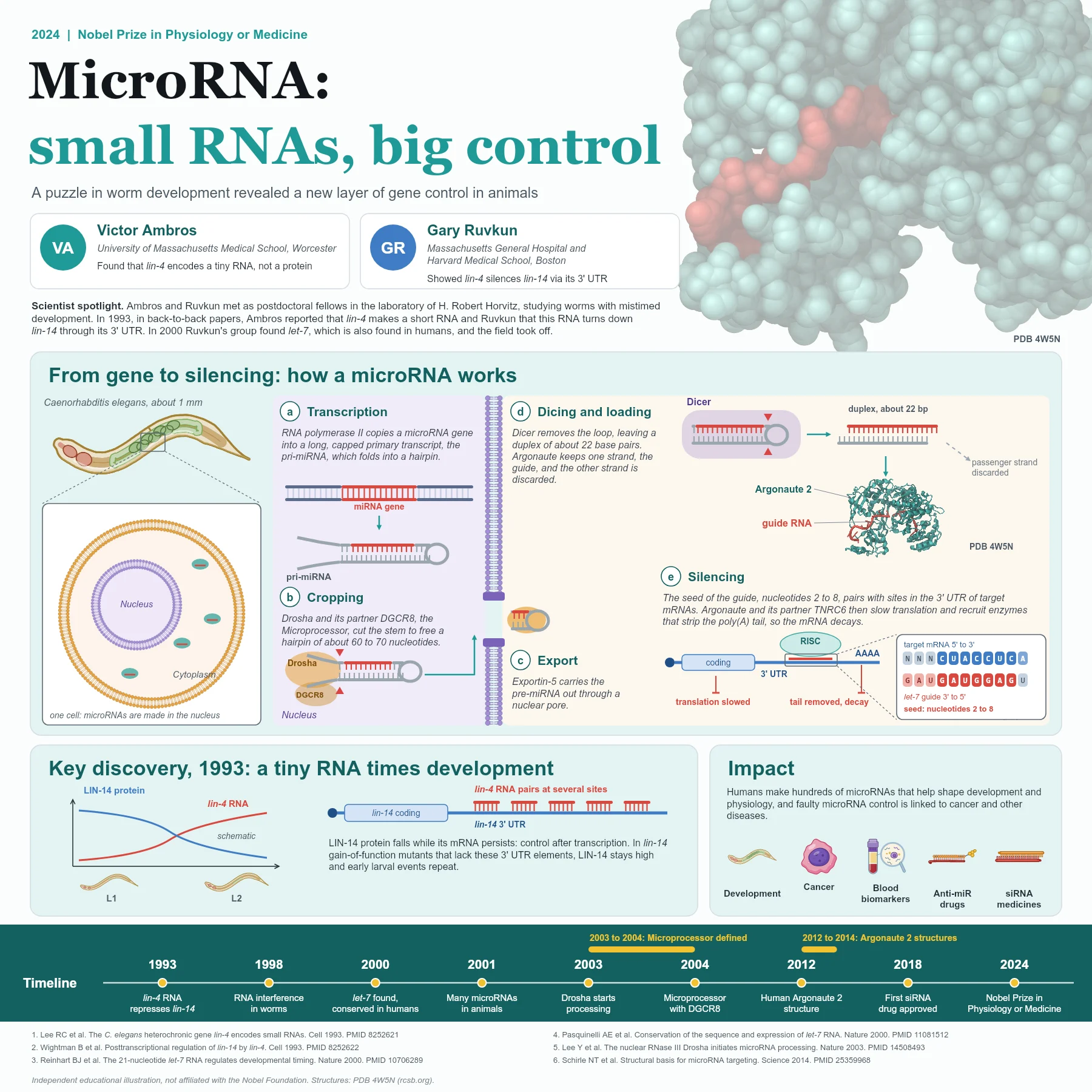
The story began with the tiny roundworm Caenorhabditis elegans, about one millimetre long, whose cells divide in a fixed and well mapped pattern. Mutant worms with timing defects, known as heterochronic mutants, either repeat early larval events or skip ahead. Two of the genes involved were lin-4 and lin-14, and genetics showed that lin-4 somehow turns lin-14 down.
The discovery story
Victor Ambros and Gary Ruvkun had both worked as postdoctoral fellows in the laboratory of H. Robert Horvitz, and each went on to study one side of the puzzle. Ambros and his colleagues cloned lin-4 and found something unexpected: the gene did not encode a protein at all. Instead it produced short RNAs, the shortest about 22 nucleotides long, with sequences partly complementary to lin-14.
Ruvkun's group studied how lin-14 is regulated. They found that the amount of LIN-14 protein falls during early larval development while its messenger RNA stays, so the control happens after transcription. The key element lay in the 3' untranslated region (3' UTR) of the lin-14 mRNA, and gain-of-function lin-14 mutants that lack parts of this region keep LIN-14 high. When the two researchers compared sequences, the lin-4 RNA matched several sites in that 3' UTR. The two papers appeared back to back in Cell in 1993.
For several years this looked like a worm curiosity. That changed in 2000, when Ruvkun's group found a second small regulatory RNA, let-7, and showed soon after with colleagues that its sequence is conserved across animals, including humans. Within a year, many such RNAs had been found in worms, flies and human cells, and they were named microRNAs.
How a microRNA works
MicroRNA genes are transcribed by RNA polymerase II into long primary transcripts, pri-miRNAs, which fold into hairpins. In the nucleus the Microprocessor, made of the enzyme Drosha and its partner DGCR8, crops each hairpin to a precursor of about 60 to 70 nucleotides. Exportin-5 carries this pre-miRNA through a nuclear pore into the cytoplasm.
There, the enzyme Dicer removes the loop and leaves a short double-stranded RNA of about 22 base pairs. One strand, the guide, is loaded into an Argonaute protein, and the other is discarded. Crystal structures of human Argonaute 2 with a bound guide RNA, such as PDB entry 4W5N, show how the protein holds the guide and presents part of it for pairing.
The most important part of the guide is its seed, nucleotides 2 to 8. The seed pairs with matching sites, usually in the 3' UTR of target mRNAs. Argonaute and its partner protein TNRC6 (GW182 in flies) then reduce translation and recruit enzymes that shorten the poly(A) tail, so the mRNA is degraded. Because the seed is short, one microRNA can regulate many different genes.
From discovery to applications
Humans make hundreds of microRNAs, and they help shape development, the immune system, the heart and the brain. Faulty microRNA control is linked to cancer and other diseases, and stable microRNAs circulating in blood are studied as biomarkers. Chemically modified oligonucleotides that block a microRNA, often called anti-miRs, have been tested as experimental drugs.
MicroRNAs share their machinery with RNA interference, discovered in worms in 1998, in which small interfering RNAs guide Argonaute to cut a fully matched target. That related pathway led to a new class of medicines, and the first siRNA drug was approved in 2018.
Why it matters today
The discovery of microRNAs changed how biologists think about genomes. Large parts of the genome produce RNAs that never become proteins, and some of them act as precise regulators. A puzzle about the timing of larval stages in a small worm opened a field that now spans plants, animals and medicine.
For students, the lesson is also about method. Both laureates relied on careful genetics in a simple animal, on cloning genes whose function was unknown, and on comparing sequences across species. The answer came not from a search for small RNAs but from the stubborn wish to understand one developmental switch. Today microRNA research spans basic biology, diagnostics and drug design, and the 1993 papers remain a model of how two complementary lines of evidence can solve a puzzle together.
Key takeaways
- lin-4, the first microRNA, was found in 1993 in the worm C. elegans and represses lin-14 through its 3' UTR.
- let-7, found in 2000, is conserved across animals including humans, which showed microRNAs are widespread.
- Drosha and DGCR8 crop pri-miRNAs in the nucleus; Exportin-5 exports them; Dicer and Argonaute finish the job in the cytoplasm.
- The seed region, nucleotides 2 to 8, guides Argonaute to 3' UTR sites and leads to repression and mRNA decay.
- MicroRNAs are studied as disease markers and drug targets, and related siRNA medicines are now approved.
References
- Lee RC, Feinbaum RL, Ambros V. The C. elegans heterochronic gene lin-4 encodes small RNAs with antisense complementarity to lin-14. Cell. 1993;75(5):843-54. PubMed 8252621
- Wightman B, Ha I, Ruvkun G. Posttranscriptional regulation of the heterochronic gene lin-14 by lin-4 mediates temporal pattern formation in C. elegans. Cell. 1993;75(5):855-62. PubMed 8252622
- Reinhart BJ, Slack FJ, Basson M, et al. The 21-nucleotide let-7 RNA regulates developmental timing in Caenorhabditis elegans. Nature. 2000;403(6772):901-6. PubMed 10706289
- Pasquinelli AE, Reinhart BJ, Slack F, et al. Conservation of the sequence and temporal expression of let-7 heterochronic regulatory RNA. Nature. 2000;408(6808):86-9. PubMed 11081512
- Lee Y, Ahn C, Han J, et al. The nuclear RNase III Drosha initiates microRNA processing. Nature. 2003;425(6956):415-9. PubMed 14508493
- Gregory RI, Yan KP, Amuthan G, et al. The Microprocessor complex mediates the genesis of microRNAs. Nature. 2004;432(7014):235-40. PubMed 15531877
- Schirle NT, Sheu-Gruttadauria J, MacRae IJ. Structural basis for microRNA targeting. Science. 2014;346(6209):608-13. PubMed 25359968
An independent educational article and illustration by BioGein, not affiliated with or endorsed by the Nobel Foundation. Protein structures are rendered from Protein Data Bank entries (rcsb.org).