The question: why did synthetic mRNA fail as a medicine?
Messenger RNA carries the instructions for making proteins from DNA to the ribosomes. Since its discovery in 1961, researchers have imagined using mRNA as a drug: deliver the right message and let the body's own cells make a therapeutic protein or a vaccine antigen. In 1990 a study showed that mRNA injected directly into mouse muscle was translated into protein, proving the principle.
For years, however, mRNA made in the laboratory by in vitro transcription had two serious problems. It was unstable and gave modest amounts of protein, and it provoked strong inflammation. Cells treated with it released cytokines and switched on antiviral defences. That made mRNA hard to use for repeated dosing and raised safety concerns for vaccines.
The discovery story: a clue in the chemistry of natural RNA
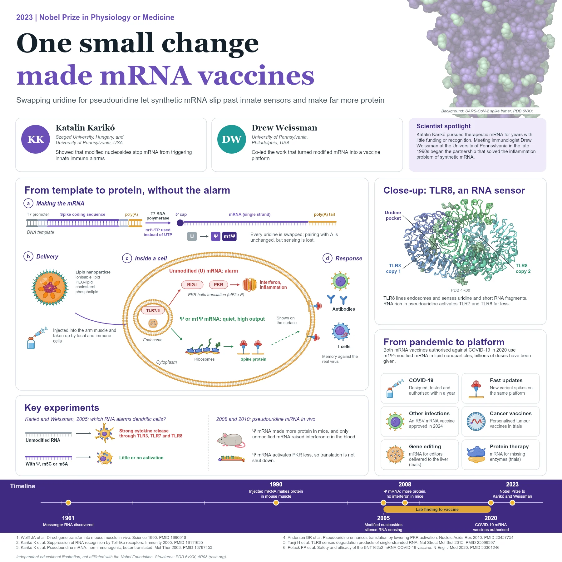
Katalin Karikó, a biochemist, had worked on therapeutic mRNA for years with little funding. At the University of Pennsylvania in the late 1990s she began working with the immunologist Drew Weissman, who wanted to use mRNA to make vaccines. They noticed that dendritic cells reacted strongly to in vitro transcribed mRNA, but much less to RNA from mammalian cells.
Natural RNA is chemically decorated. Many of its nucleosides carry modifications such as methyl groups, and transfer RNA is especially rich in pseudouridine, an isomer of uridine. Karikó and Weissman asked whether these modifications explained the difference. In 2005 they reported that RNA activated the innate sensors TLR3, TLR7 and TLR8, but that incorporating modified nucleosides such as pseudouridine, 5-methylcytidine or N6-methyladenosine abolished this activation. Dendritic cells exposed to modified RNA released far fewer cytokines.
In 2008 they showed that pseudouridine-containing mRNA was translated more efficiently in cells and in mice, and that only unmodified mRNA raised interferon-alpha in the blood. In 2010 they found one reason for the higher protein output: unmodified mRNA activates the kinase PKR, which phosphorylates the translation factor eIF2-alpha and shuts protein synthesis down, whereas pseudouridine mRNA activates PKR much less.
How a modern mRNA vaccine works
Production starts from a DNA template that carries a promoter for the bacteriophage T7 RNA polymerase, untranslated regions, the coding sequence of the antigen and a template for a poly(A) tail. During in vitro transcription, uridine triphosphate is replaced by a modified version, today usually N1-methylpseudouridine triphosphate, so every uridine in the mRNA is swapped. The modified base still pairs with adenosine, so the genetic code is unchanged. A 5' cap is added for efficient translation.
The mRNA is packaged in lipid nanoparticles made of an ionisable lipid, a PEG-lipid, cholesterol and a phospholipid. After injection into the arm muscle, cells near the injection site and immune cells in draining lymph nodes take up the particles by endocytosis. A fraction of the mRNA escapes from endosomes into the cytoplasm, where ribosomes translate it. Because the modified mRNA triggers TLR7, TLR8 and other sensors far less, translation continues and cells make abundant antigen.
For COVID-19 vaccines the antigen is the SARS-CoV-2 spike protein, which is displayed on cells and presented to the immune system. B cells make neutralising antibodies and T cells are primed, giving memory that protects against the real virus.
From discovery to applications
When the SARS-CoV-2 genome was published in January 2020, nucleoside-modified mRNA in lipid nanoparticles was ready to be adapted. Two mRNA vaccines encoding the spike protein were designed, tested in large trials and authorised in December 2020. In one trial with tens of thousands of participants, the vaccine showed 95% efficacy against symptomatic COVID-19. Billions of doses have since been given, and updated vaccines for new variants use the same platform.
The technology is now being applied to other infections, including an mRNA vaccine against respiratory syncytial virus approved in 2024, to personalised cancer vaccines, to protein replacement for rare diseases and to delivering gene editors to the liver. Many of these uses remain in clinical trials.
Why it matters today
The key insight was chemical and immunological at once: innate immunity distinguishes self RNA from foreign RNA partly by its nucleoside modifications, and synthetic RNA can be made to look like self. That insight turned mRNA from a promising but inflammatory molecule into a practical platform that can be redesigned in weeks. It is also a reminder that persistent, curiosity-driven work, often with little recognition, can prepare the ground for a sudden global need.
Several other advances had to come together. Lipid nanoparticles, first developed to deliver small interfering RNA, protect the fragile mRNA and help it into cells. Structural work on coronavirus spike proteins showed how to lock the spike in its prefusion shape, the form that neutralising antibodies recognise best. Improved manufacturing and purification removed contaminating double-stranded RNA, another trigger of innate sensors. Nucleoside modification was the foundation on which these pieces could be combined into a vaccine that is potent, well tolerated and quick to update.
Key takeaways
- Unmodified synthetic mRNA activates innate sensors such as TLR3, TLR7, TLR8 and PKR.
- Karikó and Weissman showed in 2005 that nucleoside modifications such as pseudouridine abolish TLR activation.
- Pseudouridine mRNA is translated more efficiently, partly because it activates PKR less.
- COVID-19 mRNA vaccines use N1-methylpseudouridine mRNA in lipid nanoparticles encoding the spike protein.
- The platform is being extended to other vaccines, cancer and protein replacement.
References
- Wolff JA, Malone RW, Williams P, et al. Direct gene transfer into mouse muscle in vivo. Science. 1990;247(4949 Pt 1):1465-8. PubMed 1690918
- Karikó K, Buckstein M, Ni H, Weissman D. Suppression of RNA recognition by Toll-like receptors: the impact of nucleoside modification and the evolutionary origin of RNA. Immunity. 2005;23(2):165-75. PubMed 16111635
- Karikó K, Muramatsu H, Welsh FA, et al. Incorporation of pseudouridine into mRNA yields superior nonimmunogenic vector with increased translational capacity and biological stability. Mol Ther. 2008;16(11):1833-40. PubMed 18797453
- Anderson BR, Muramatsu H, Nallagatla SR, et al. Incorporation of pseudouridine into mRNA enhances translation by diminishing PKR activation. Nucleic Acids Res. 2010;38(17):5884-92. PubMed 20457754
- Tanji H, Ohto U, Shibata T, et al. Toll-like receptor 8 senses degradation products of single-stranded RNA. Nat Struct Mol Biol. 2015;22(2):109-15. PubMed 25599397
- Walls AC, Park YJ, Tortorici MA, et al. Structure, function, and antigenicity of the SARS-CoV-2 spike glycoprotein. Cell. 2020;181(2):281-292.e6. PubMed 32155444
- Polack FP, Thomas SJ, Kitchin N, et al. Safety and efficacy of the BNT162b2 mRNA Covid-19 vaccine. N Engl J Med. 2020;383(27):2603-2615. PubMed 33301246
An independent educational article and illustration by BioGein, not affiliated with or endorsed by the Nobel Foundation. Protein structures are rendered from Protein Data Bank entries (rcsb.org).