The question: how does a cell know how much oxygen it has?
Animals depend on oxygen to make most of their energy, yet the supply changes all the time. Climb a mountain, lose blood, exercise hard or develop a tumour that outgrows its blood vessels, and some cells find themselves short of oxygen. The body responds in many ways: the kidney releases more of the hormone erythropoietin (EPO), the bone marrow makes more red blood cells, new blood vessels sprout and cells switch towards glycolysis, which makes ATP without oxygen.
For decades it was unclear how these responses were triggered. Was there a dedicated oxygen sensor in a few specialised cells, or could every cell measure oxygen for itself? The work honoured in 2019 answered this by uncovering a molecular switch that operates in almost every cell of the body.
The discovery story
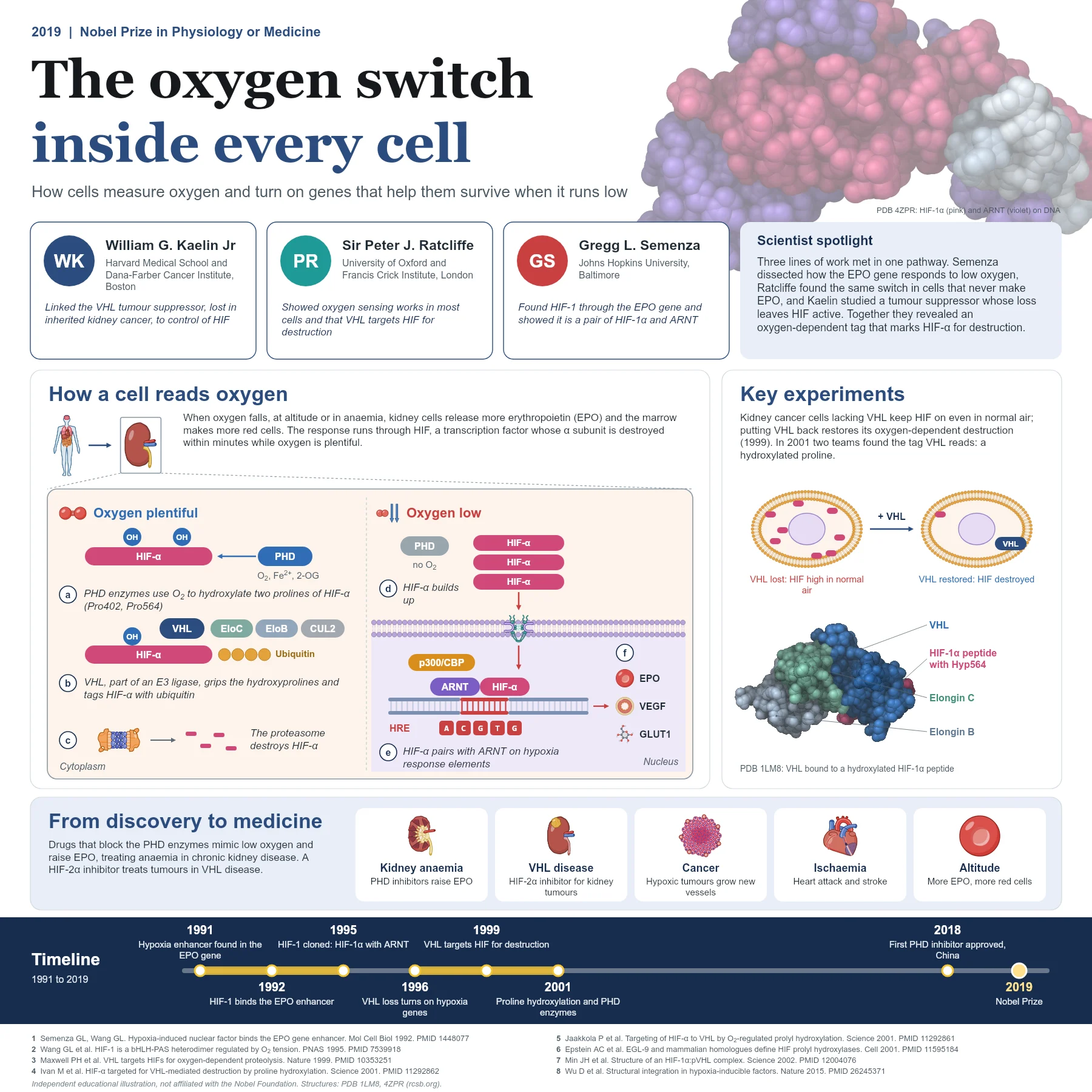
Gregg Semenza approached the problem through the EPO gene. His group mapped a short enhancer next to the gene that switched on transcription when oxygen fell, and in 1992 showed that a nuclear factor, induced by hypoxia, bound to it. They called it hypoxia-inducible factor 1 (HIF-1). In 1995 they purified and cloned it and found that it consists of two partners: HIF-1α, which responds to oxygen, and a constant partner already known as ARNT, also called HIF-1β.
Peter Ratcliffe, studying the same EPO control system, found that the oxygen-sensing machinery is not limited to kidney or liver cells that make EPO. It works in nearly all cell types, which meant that oxygen sensing was a general property of cells.
William Kaelin came to the field from cancer research. People with von Hippel-Lindau (VHL) disease inherit a faulty copy of the VHL gene and develop tumours, including kidney cancers, that are unusually rich in blood vessels. Kaelin showed in 1996 that cells lacking functional VHL switch on hypoxia-regulated genes even in normal air. In 1999 Ratcliffe and colleagues reported that VHL binds HIF-α and is needed for its oxygen-dependent destruction. Finally, in 2001, the Kaelin and Ratcliffe groups showed that VHL recognises HIF-α only after specific proline residues have been hydroxylated, a modification that requires molecular oxygen.
How the mechanism works
When oxygen is plentiful, enzymes called prolyl hydroxylases (PHDs) use O2, iron and 2-oxoglutarate to attach hydroxyl groups to two prolines in HIF-α (Pro402 and Pro564 in human HIF-1α). The hydroxylated prolines fit into a pocket of the VHL protein, which is part of an E3 ubiquitin ligase together with Elongin B, Elongin C and Cullin 2. The ligase decorates HIF-α with ubiquitin chains and the proteasome destroys it within minutes. The structure of VHL bound to a hydroxylated HIF-1α peptide (PDB 1LM8) shows exactly how the hydroxyproline is recognised.
When oxygen falls, the PHDs slow down because they lack their co-substrate. HIF-α escapes hydroxylation, accumulates and moves into the nucleus. There it pairs with ARNT and binds short DNA sequences called hypoxia response elements, with the core sequence RCGTG, together with co-activators such as p300 and CBP. Hundreds of target genes follow, including EPO for red cell production, VEGF for new blood vessels and genes for glucose transport and glycolysis such as GLUT1. A related enzyme, FIH, adds a second oxygen-dependent control by blocking the co-activator interaction.
Because HIF-α is made continuously, its level is set mainly by how fast it is destroyed. The hydroxylases have a relatively low affinity for oxygen, so their activity falls across the physiological range and they behave as true sensors. The response is graded rather than all or nothing. HIF-2α, a close relative of HIF-1α that uses the same partner, controls an overlapping set of genes and is the main driver of EPO production in the adult kidney.
From discovery to medicine
Because the PHD enzymes are the sensor, blocking them mimics low oxygen. Oral PHD inhibitors raise the body’s own EPO and are used to treat anaemia in chronic kidney disease; the first, roxadustat, was approved in China in 2018. In the opposite direction, a drug that blocks HIF-2α, belzutifan, treats kidney and other tumours in people with VHL disease.
The HIF system also shapes how tumours grow and resist therapy, how the heart and brain cope with interrupted blood flow, and how people adapt to life at high altitude, making it a central pathway in physiology and disease.
Why it matters today
The oxygen switch is a striking example of basic curiosity becoming medicine within two decades. A question about one hormone gene led to a general sensing mechanism, a family of enzymes, a tumour suppressor’s function and new classes of drugs. Structural studies, including the HIF-1α and ARNT complex bound to DNA (PDB 4ZPR), continue to guide the design of molecules that tune this pathway with more precision.
Key takeaways
- HIF-α is made constantly but destroyed within minutes while oxygen is available.
- PHD enzymes use oxygen to hydroxylate HIF-α prolines, creating the tag that VHL recognises.
- In hypoxia, HIF-α pairs with ARNT on hypoxia response elements and activates EPO, VEGF and glycolytic genes.
- PHD inhibitors treat anaemia of kidney disease, and a HIF-2α inhibitor treats tumours in VHL disease.
References
- Semenza GL, Wang GL. A nuclear factor induced by hypoxia via de novo protein synthesis binds to the human erythropoietin gene enhancer at a site required for transcriptional activation. Mol Cell Biol. 1992;12(12):5447-54. PubMed 1448077
- Wang GL, Jiang BH, Rue EA, et al. Hypoxia-inducible factor 1 is a basic-helix-loop-helix-PAS heterodimer regulated by cellular O2 tension. Proc Natl Acad Sci U S A. 1995;92(12):5510-4. PubMed 7539918
- Iliopoulos O, Levy AP, Jiang C, et al. Negative regulation of hypoxia-inducible genes by the von Hippel-Lindau protein. Proc Natl Acad Sci U S A. 1996;93(20):10595-9. PubMed 8855223
- Maxwell PH, Wiesener MS, Chang GW, et al. The tumour suppressor protein VHL targets hypoxia-inducible factors for oxygen-dependent proteolysis. Nature. 1999;399(6733):271-5. PubMed 10353251
- Ivan M, Kondo K, Yang H, et al. HIFalpha targeted for VHL-mediated destruction by proline hydroxylation: implications for O2 sensing. Science. 2001;292(5516):464-8. PubMed 11292862
- Jaakkola P, Mole DR, Tian YM, et al. Targeting of HIF-alpha to the von Hippel-Lindau ubiquitylation complex by O2-regulated prolyl hydroxylation. Science. 2001;292(5516):468-72. PubMed 11292861
- Min JH, Yang H, Ivan M, et al. Structure of an HIF-1alpha -pVHL complex: hydroxyproline recognition in signaling. Science. 2002;296(5574):1886-9. PubMed 12004076
- Wu D, Potluri N, Lu J, et al. Structural integration in hypoxia-inducible factors. Nature. 2015;524(7565):303-8. PubMed 26245371
An independent educational article and illustration by BioGein, not affiliated with or endorsed by the Nobel Foundation. Protein structures are rendered from Protein Data Bank entries (rcsb.org).