The question: why does the immune system tolerate cancer?
Tumour cells carry mutated and abnormally expressed proteins that T cells can recognise. Yet tumours often grow in the presence of tumour-specific T cells. For most of the twentieth century, attempts to turn the immune system against cancer, from bacterial toxins to cytokines, gave occasional dramatic responses but rarely lasting benefit for most patients.
A different idea emerged from basic research on how T cells are switched on and off. T cell activation needs more than antigen recognition: it also depends on costimulatory and inhibitory receptors that act like an accelerator and brakes. If tumours exploit the brakes, then releasing them might restore an attack that is already primed.
The discovery story: two brakes, two laboratories
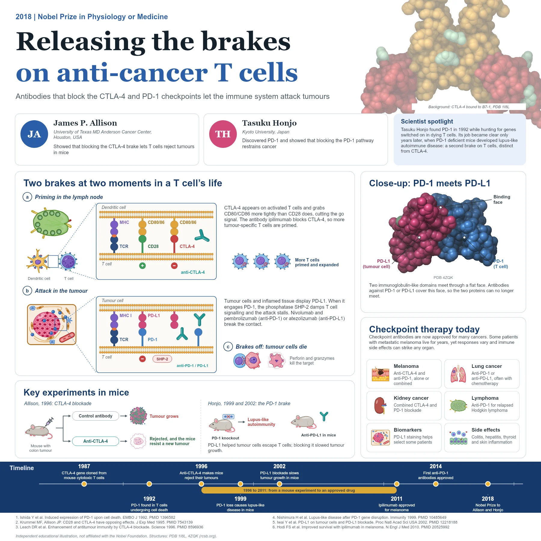
CTLA-4 was cloned in 1987 as a new member of the immunoglobulin superfamily expressed by activated T cells. It resembles CD28, the receptor that delivers the main costimulatory signal when it binds CD80 or CD86 on antigen-presenting cells. In 1995, Matthew Krummel and James Allison showed that CTLA-4 and CD28 have opposing effects: engaging CTLA-4 inhibits T cell proliferation and IL-2 production. Other groups reached similar conclusions, and mice lacking CTLA-4 develop fatal lymphoproliferation.
Allison reasoned that blocking this brake could help T cells reject tumours. In 1996, his group reported that antibodies against CTLA-4 caused mice to reject transplanted tumours, including established ones, and that treated mice resisted a second tumour challenge. This experiment launched the development of ipilimumab.
In Kyoto, Tasuku Honjo's group had identified PD-1 in 1992 while searching for genes switched on in T cells undergoing programmed cell death. Its function only became clear later: in 1999 they showed that PD-1 deficient mice develop lupus-like arthritis and glomerulonephritis, revealing PD-1 as a negative regulator of immune responses. With collaborators they identified its ligand, PD-L1, in 2000. In 2002, Honjo's group showed that PD-L1 on tumour cells protects them from cytotoxic T cells and that blocking PD-L1 restrained tumour growth in mice.
How checkpoint blockade works
The two brakes act at different moments. CTLA-4 works mainly during priming in lymph nodes. When a naive T cell meets a dendritic cell presenting its antigen, CD28 receives the go signal from CD80 and CD86. Activated T cells then raise CTLA-4, which binds the same ligands with higher affinity and limits further activation. An anti-CTLA-4 antibody such as ipilimumab blocks this competition, so more tumour-specific T cells are primed and expanded.
PD-1 acts mainly later, in tissues and tumours. Activated and chronically stimulated T cells express PD-1. When it engages PD-L1 on tumour cells or inflamed tissue, it recruits the phosphatase SHP-2, which damps signals from the T cell receptor and CD28, and the attack stalls. Antibodies against PD-1 (nivolumab, pembrolizumab) or PD-L1 (atezolizumab and others) cover the flat binding face seen in the crystal structure of the PD-1/PD-L1 complex, so the two proteins cannot meet and T cells can resume killing with perforin and granzymes.
From discovery to the clinic
In 2010 a randomised trial showed that ipilimumab improved overall survival in patients with metastatic melanoma, a disease that had few effective treatments. Ipilimumab was approved in 2011. The first anti-PD-1 antibodies were approved in 2014 and proved active in a broad range of cancers, with fewer severe side effects than anti-CTLA-4.
Today checkpoint inhibitors are used in melanoma, lung, kidney, bladder and head and neck cancers, Hodgkin lymphoma and tumours with mismatch repair deficiency, among others, sometimes combined with each other or with chemotherapy. A minority of patients with advanced disease achieve long-lasting control. Because the brakes also protect healthy tissue, side effects are immune-related and can affect the gut, liver, skin, lungs and endocrine glands.
Why it matters today
Checkpoint blockade established immunotherapy as a pillar of cancer treatment alongside surgery, radiation, chemotherapy and targeted drugs. It also raised new questions that drive research: why only some patients respond, how to predict response with markers such as PD-L1 staining or tumour mutational burden, and how to combine checkpoint drugs with vaccines, cell therapies and other treatments. Both discoveries began as curiosity about how T cells are regulated, not as cancer projects.
The two brakes also explain why the side effects look the way they do. Mice lacking CTLA-4 die from uncontrolled T cell expansion, while mice lacking PD-1 develop slower, organ-specific autoimmunity. In patients, anti-CTLA-4 treatment tends to cause more frequent and severe inflammation of the gut and skin than anti-PD-1 treatment, and combining the two increases both benefit and toxicity. Managing these immune-related adverse events, usually with corticosteroids, has become a new skill for oncologists, and it is a direct reminder that checkpoints exist to protect the body from its own immune system.
Key takeaways
- CTLA-4 and PD-1 are inhibitory receptors that act as brakes on T cells.
- Allison showed in 1996 that blocking CTLA-4 lets mice reject tumours.
- Honjo discovered PD-1 in 1992 and showed that it restrains immune responses and protects tumours through PD-L1.
- Anti-CTLA-4 acts mainly during priming; anti-PD-1 and anti-PD-L1 act mainly in the tumour.
- Checkpoint inhibitors are approved for many cancers but cause immune-related side effects.
References
- Brunet JF, Denizot F, Luciani MF, et al. A new member of the immunoglobulin superfamily: CTLA-4. Nature. 1987;328(6127):267-70. PubMed 3496540
- Ishida Y, Agata Y, Shibahara K, Honjo T. Induced expression of PD-1, a novel member of the immunoglobulin gene superfamily, upon programmed cell death. EMBO J. 1992;11(11):3887-95. PubMed 1396582
- Krummel MF, Allison JP. CD28 and CTLA-4 have opposing effects on the response of T cells to stimulation. J Exp Med. 1995;182(2):459-65. PubMed 7543139
- Leach DR, Krummel MF, Allison JP. Enhancement of antitumor immunity by CTLA-4 blockade. Science. 1996;271(5256):1734-6. PubMed 8596936
- Nishimura H, Nose M, Hiai H, Minato N, Honjo T. Development of lupus-like autoimmune diseases by disruption of the PD-1 gene encoding an ITIM motif-carrying immunoreceptor. Immunity. 1999;11(2):141-51. PubMed 10485649
- Iwai Y, Ishida M, Tanaka Y, Okazaki T, Honjo T, Minato N. Involvement of PD-L1 on tumor cells in the escape from host immune system and tumor immunotherapy by PD-L1 blockade. Proc Natl Acad Sci U S A. 2002;99(19):12293-7. PubMed 12218188
- Hodi FS, O'Day SJ, McDermott DF, et al. Improved survival with ipilimumab in patients with metastatic melanoma. N Engl J Med. 2010;363(8):711-23. PubMed 20525992
- Zak KM, Kitel R, Przetocka S, et al. Structure of the complex of human programmed death 1, PD-1, and its ligand PD-L1. Structure. 2015;23(12):2341-8. PubMed 26602187
An independent educational article and illustration by BioGein, not affiliated with or endorsed by the Nobel Foundation. Protein structures are rendered from Protein Data Bank entries (rcsb.org).