The question: can we borrow evolution's method?
Natural evolution improves proteins by a simple recipe: variation in genes, selection of the variants that work best, and repetition over many generations. Designing better enzymes or antibodies from first principles is hard because we still cannot reliably predict which changes in a sequence will improve function.
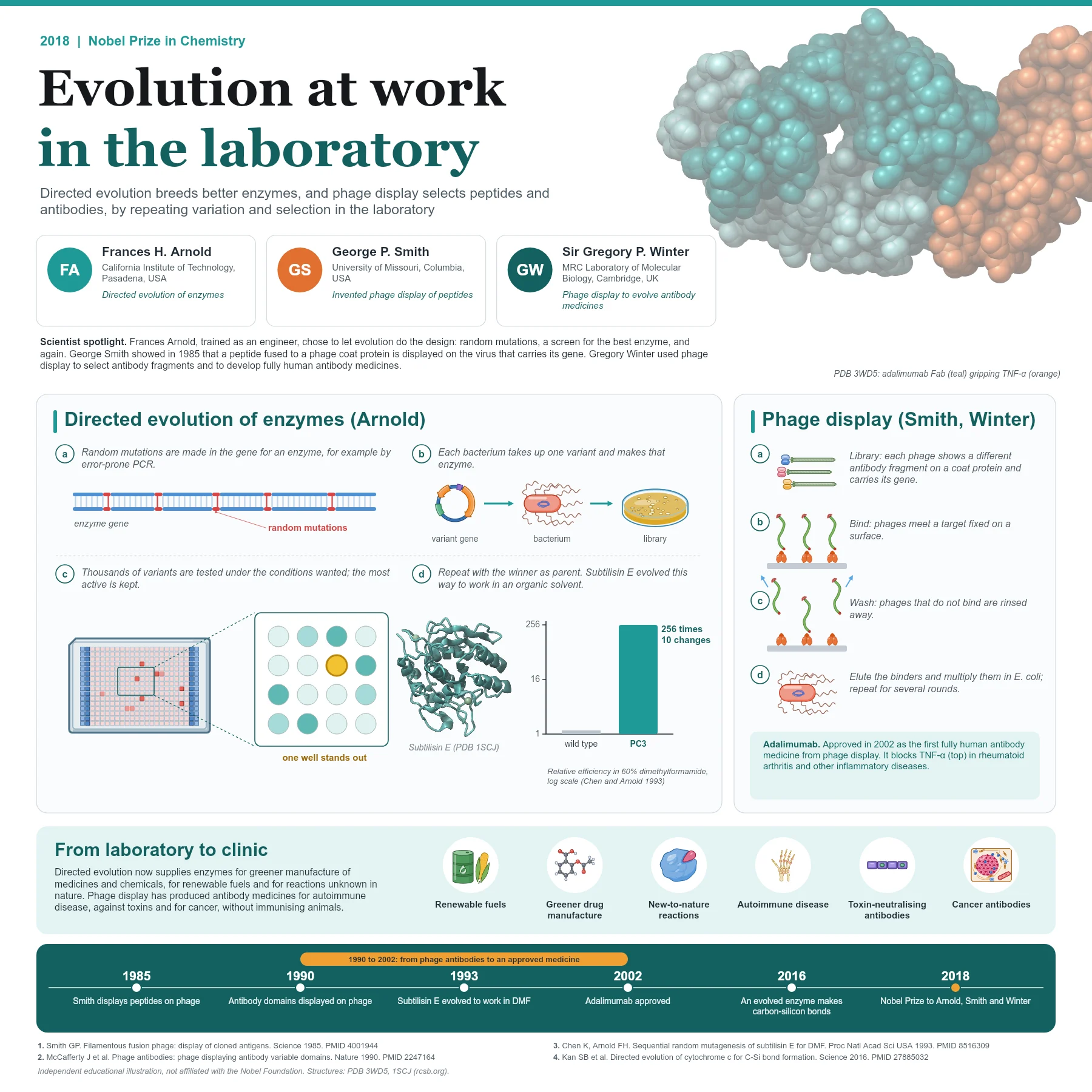
The 2018 Nobel Prize in Chemistry recognised scientists who put evolution's recipe to work in the laboratory. One half went to Frances H. Arnold for the directed evolution of enzymes, and the other half jointly to George P. Smith and Sir Gregory P. Winter for the phage display of peptides and antibodies.
Directed evolution of enzymes
Arnold, trained as an engineer, decided to stop trying to design enzymes rationally and to let random variation and screening do the work. In a landmark 1993 study, she and Keqing Chen took subtilisin E, a protease that normally works in water, and set out to make it work in a high concentration of the organic solvent dimethylformamide.
They introduced random mutations into the gene, expressed the variants in bacteria and screened them for activity in the solvent. The best variant became the parent for the next round. After sequential rounds, the variant PC3 carried ten amino acid substitutions and hydrolysed a peptide substrate 256 times more efficiently than the wild-type enzyme in 60% dimethylformamide. Many of the substitutions lay in surface loops near the active site, places that no one would have picked in advance.
Directed evolution has since been used to create enzymes with entirely new chemistry. In 2016 Arnold's group evolved a bacterial cytochrome c to form carbon-silicon bonds, a reaction not known in nature.
How well the approach works depends on the test. A screen measures each variant on its own, for example by colour or fluorescence in the wells of a plate, while a selection ties survival or growth to the property wanted, so that only good variants remain. Because evolution delivers exactly what it is asked for, the test must match the property that is really needed. Arnold also showed that useful mutations found in separate variants can be brought together by recombining genes, which speeds up improvement.
Phage display of peptides and antibodies
In 1985 George Smith showed that a foreign DNA fragment inserted into gene III of a filamentous phage is expressed as part of a coat protein. The resulting fusion phage displays the foreign peptide on its surface while carrying the gene for that peptide inside. Phages that bind a target can therefore be enriched more than a thousandfold by affinity, and the binding sequence can be read from their DNA.
Gregory Winter and his colleagues in Cambridge applied the idea to antibodies. In 1990 they showed that complete antibody variable domains could be displayed on phage, that the phage bound specifically to antigen, and that rare binders, one in a million, could be isolated. Selection works in rounds called panning: the library is exposed to a target fixed on a surface, unbound phages are washed away, bound phages are eluted and multiplied in bacteria, and the cycle is repeated.
The heart of phage display is the physical link between what a molecule does and the gene that encodes it. Each phage particle carries its own gene, so binders picked out of a library of many millions can simply be multiplied and sequenced. Peptide libraries were used to map the sites that antibodies recognise, and antibody libraries could be assembled from human variable genes, which meant that human antibodies could be obtained without immunising a person or an animal.
Winter's group went further and used repeated rounds of mutation and selection on phage to improve the affinity of selected antibodies, bringing directed evolution and display together in one method.
From discovery to medicines and manufacturing
Phage display made it possible to obtain fully human antibodies without immunising animals. Adalimumab, which blocks the inflammatory signal TNF-alpha, was approved in 2002 as the first fully human antibody medicine developed with phage display and is used for rheumatoid arthritis and other inflammatory diseases. Further phage-derived antibodies neutralise toxins or treat cancer and autoimmune disease.
Directed evolution, in turn, supplies enzymes for greener manufacture of pharmaceuticals and other chemicals, replacing toxic catalysts and solvents, and for the production of renewable fuels.
Both methods also make good partners for structural and computational work. Once a selected binder or an improved enzyme is in hand, its structure and its mutations show why it works, and those lessons feed the design of the next library.
Why it matters today
Both methods share one insight: when the rules connecting sequence to function are too complex to predict, a well-designed selection can find the answer. Libraries, screens and rounds of selection are now standard tools in biotechnology, and they increasingly work together with computational design to search sequence space faster.
The two strands of the prize share one insight: when the rules linking sequence to function are too complex to predict, a well-designed selection can still find the answer. Libraries, screens and rounds of selection are now routine in biotechnology, and they increasingly work alongside computational design to search sequence space faster.
Key takeaways
- Directed evolution repeats random mutation, expression and screening to improve enzymes.
- Subtilisin E variant PC3, with ten substitutions, became 256 times more efficient in 60% dimethylformamide (1993).
- Phage display links a displayed peptide or antibody to the gene that encodes it (Smith, 1985).
- Antibody fragments were displayed on phage and rare binders isolated in 1990 (Winter's group).
- Adalimumab, approved in 2002, was the first fully human antibody medicine from phage display.
References
- Smith GP. Filamentous fusion phage: novel expression vectors that display cloned antigens on the virion surface. Science. 1985;228(4705):1315-1317. PubMed 4001944
- McCafferty J, Griffiths AD, Winter G, Chiswell DJ. Phage antibodies: filamentous phage displaying antibody variable domains. Nature. 1990;348(6301):552-554. PubMed 2247164
- Chen K, Arnold FH. Tuning the activity of an enzyme for unusual environments: sequential random mutagenesis of subtilisin E for catalysis in dimethylformamide. Proc Natl Acad Sci U S A. 1993;90(12):5618-5622. PubMed 8516309
- Kan SB, Lewis RD, Chen K, et al. Directed evolution of cytochrome c for carbon-silicon bond formation: bringing silicon to life. Science. 2016;354:1048-1051. PubMed 27885032
An independent educational article and illustration by BioGein, not affiliated with or endorsed by the Nobel Foundation. Protein structures are rendered from Protein Data Bank entries (rcsb.org).