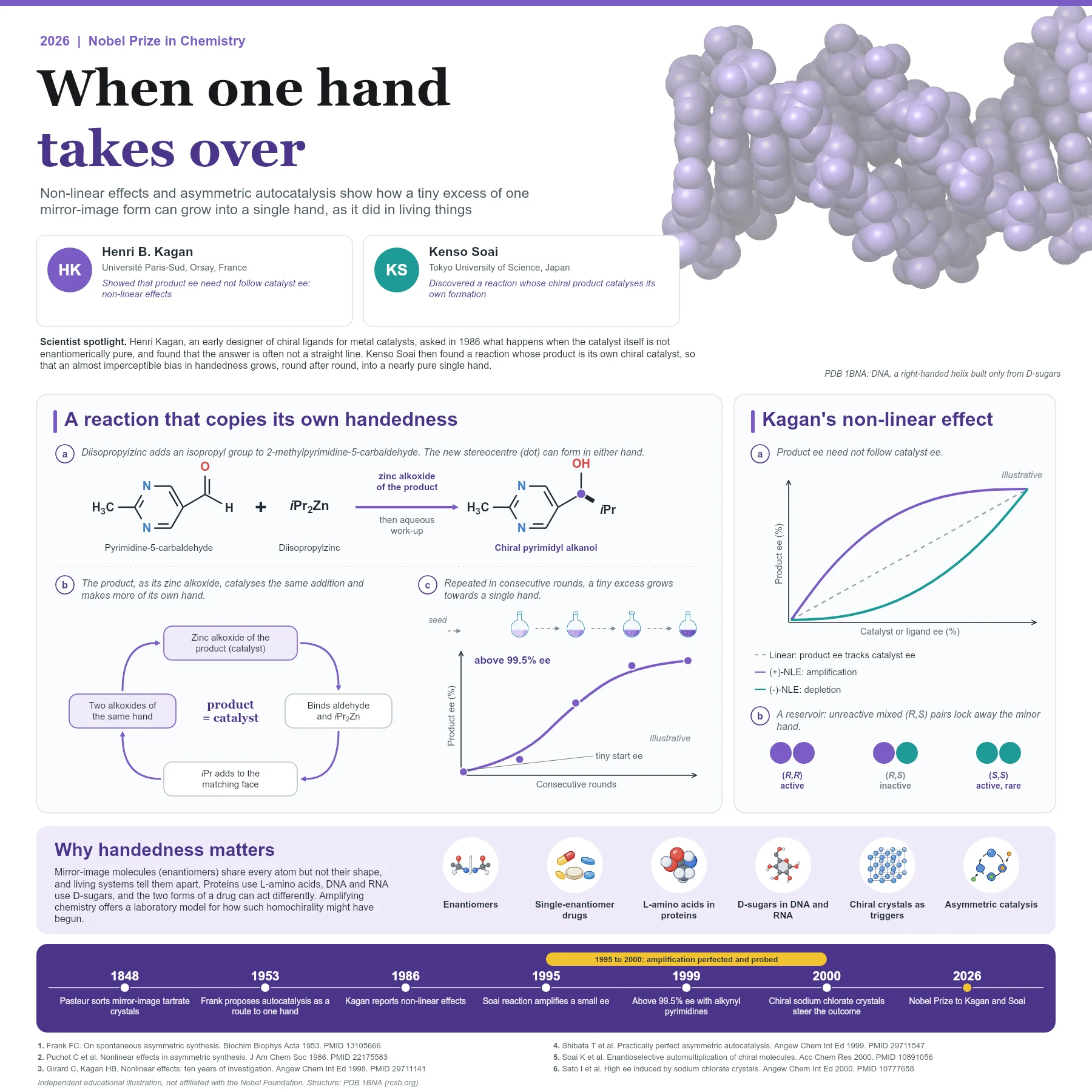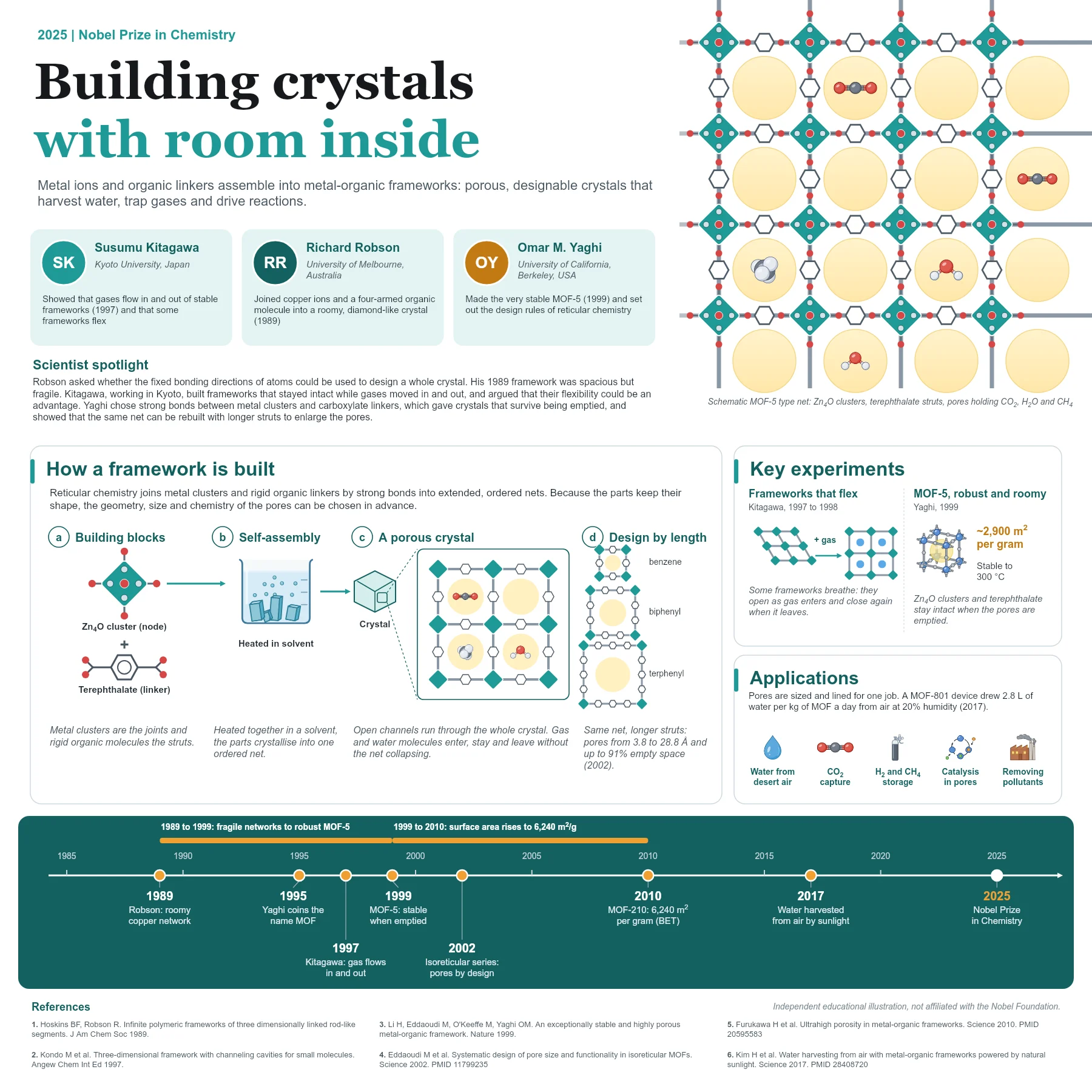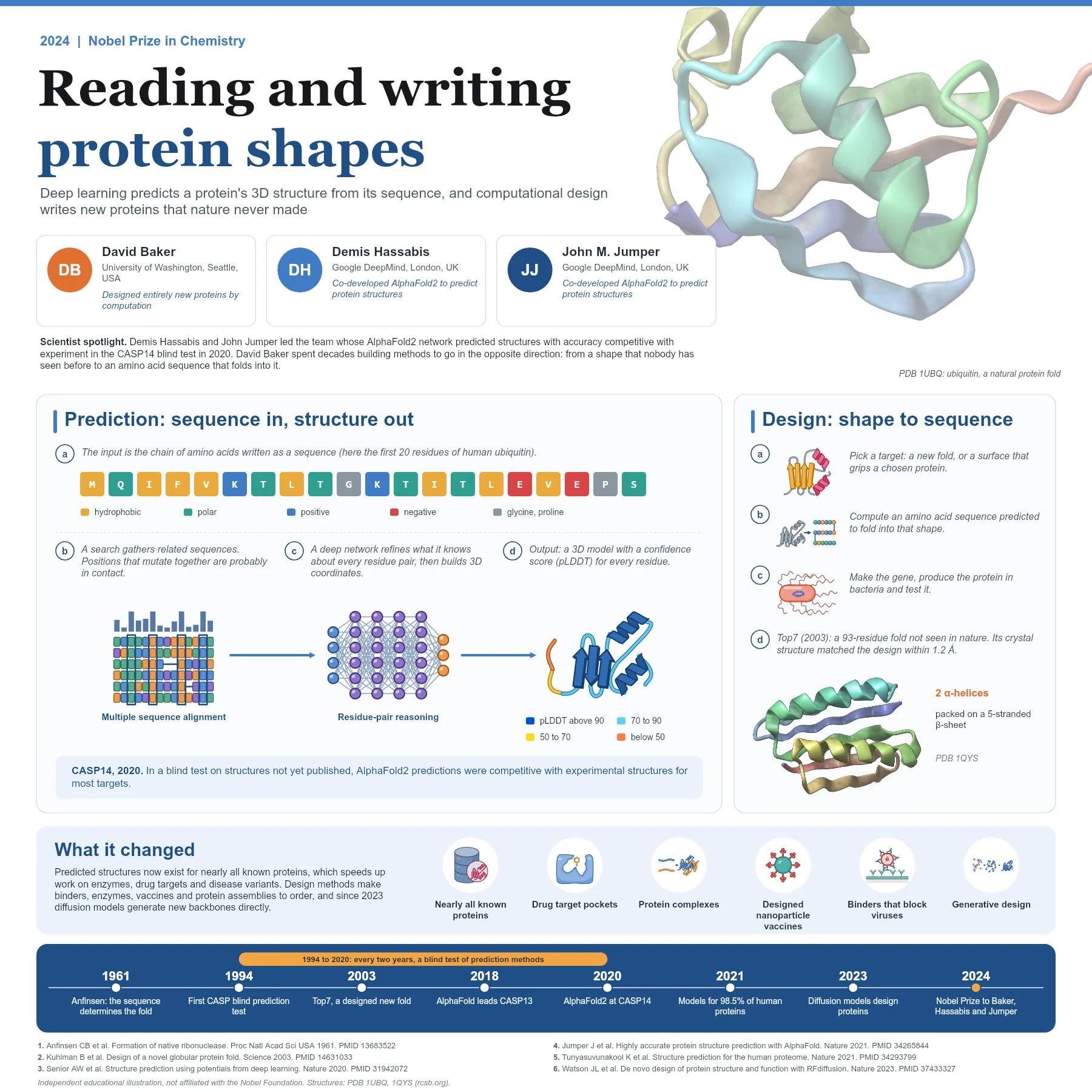
The question: can an electron microscope see a protein's atoms?
Electrons have wavelengths far shorter than the spacing between atoms, so in principle an electron microscope can resolve atomic detail. In practice biological samples posed three problems. The microscope works in a vacuum, which dries out water-based samples. The electron beam damages the molecules it images. And a single, weakly scattering protein gives a very noisy picture.
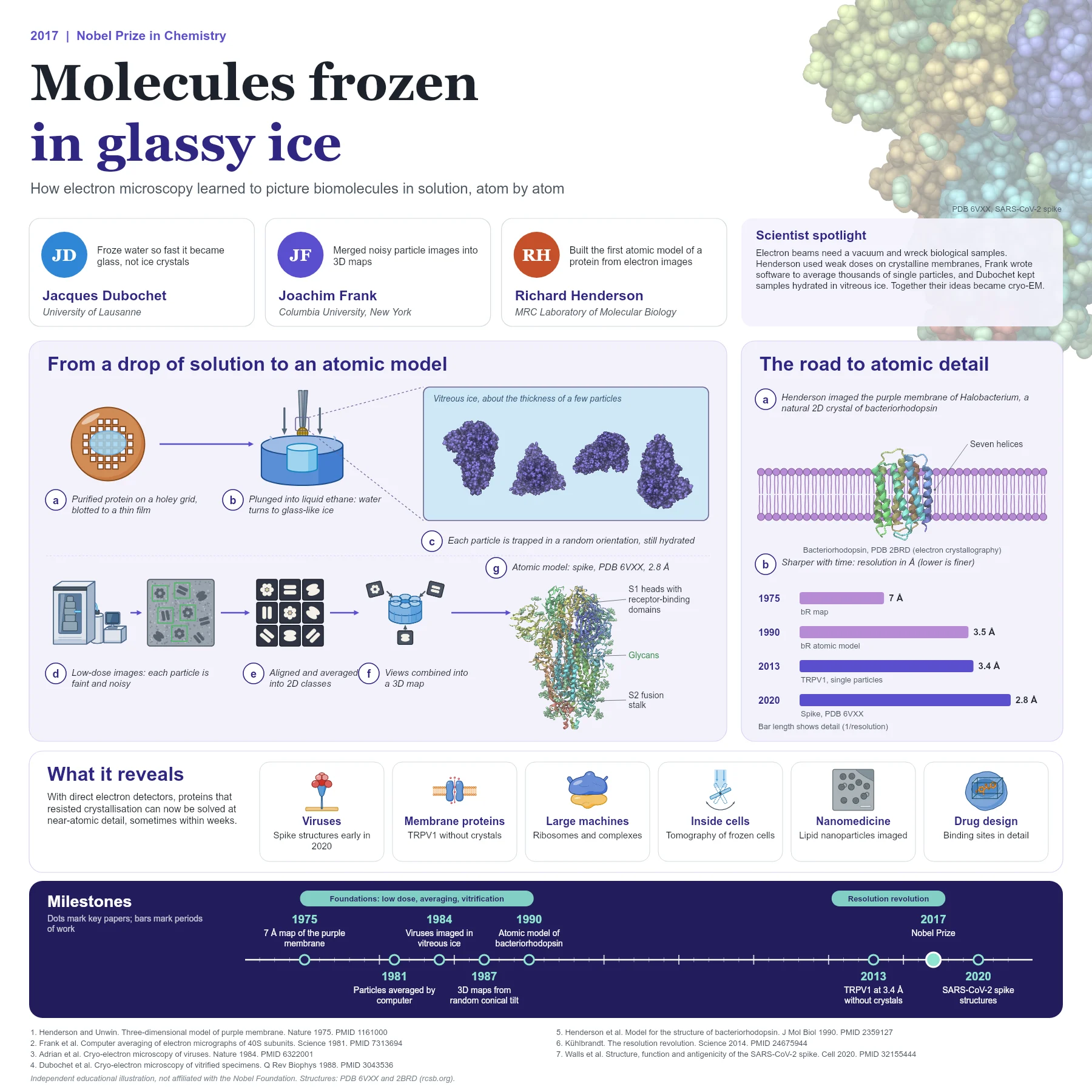
The 2017 Nobel Prize in Chemistry was awarded to Jacques Dubochet, Joachim Frank and Richard Henderson for developing cryo-electron microscopy (cryo-EM) for high-resolution structure determination of biomolecules in solution. Each solved part of the problem.
X-ray crystallography had already given many protein structures, but it needs well-ordered crystals. Many membrane proteins and large, flexible complexes never form them, which is why a method that works on molecules in solution mattered so much.
Henderson: low doses and a natural crystal
Richard Henderson worked with the purple membrane of the salt-loving archaeon Halobacterium, in which the light-driven proton pump bacteriorhodopsin forms a regular two-dimensional crystal. By using very low electron doses and averaging information across the whole crystal lattice, Henderson and Nigel Unwin obtained a three-dimensional map at 7 Å resolution in 1975. It showed seven closely packed helices crossing the membrane.
Henderson kept improving the method. In 1990 he and colleagues used electron cryo-microscopy of tilted crystals to build a complete atomic model of bacteriorhodopsin, with the retinal chromophore and the residues of the proton pathway. It proved that electron microscopy could reach atomic detail for a protein.
The 1990 model combined data from 72 images of tilted and untilted crystals with 150 electron diffraction patterns, reaching 3.5 Å resolution parallel to the membrane. Bulky aromatic side chains and the beta-ionone ring of retinal served as landmarks for building the chain.
Frank: averaging single particles
Most proteins do not form crystals. Joachim Frank developed computational methods to work with single, randomly oriented particles. In 1981 he and colleagues averaged electron images of individual ribosomal subunits by computer and obtained a clearer view than any single image.
His approach sorts thousands of noisy particle images into groups that share the same orientation, averages each group into a clean two-dimensional class, and then combines the different views into a three-dimensional map. In 1987 his group applied a random conical tilt scheme to reconstruct the 50S ribosomal subunit in three dimensions.
Applying crystallographic criteria to independent averages, Frank showed that results from single particles could be as reproducible as those from thin, stained protein crystals. This gave confidence that molecules need not be crystallised at all.
Dubochet: water turned to glass
Freezing a sample keeps it hydrated in the vacuum and reduces beam damage, but ordinary freezing forms ice crystals that scatter electrons and distort the molecules. Jacques Dubochet and his team found that a thin film of water cooled extremely fast does not crystallise and instead becomes vitreous, glass-like ice.
In 1984 they published images of unstained viruses embedded in thin vitrified films, free of the damage caused by drying or staining. The routine method is simple: a small drop of sample is placed on a holey grid, blotted to a thin film and plunged into liquid ethane cooled by liquid nitrogen.
The vitrified viral particles looked free of the damage caused by dehydration, freezing or adsorption to a support film, problems that had limited conventional electron microscopy of biological samples. Dubochet and colleagues summarised the method and its physics in a long review in 1988, which became a reference for the field.
The resolution revolution and why it matters
Around 2012 new direct electron detectors, which record movies so that sample motion can be corrected, together with better software, sharply improved image quality. In 2013 the structure of the TRPV1 ion channel was determined at 3.4 Å resolution without crystals, a milestone for membrane proteins. In 2014 a commentary named this change the resolution revolution.
Cryo-EM now solves structures of membrane proteins, large molecular machines and flexible complexes that resisted crystallisation. When SARS-CoV-2 emerged, cryo-EM structures of its spike glycoprotein, such as the 2.8 Å closed prefusion structure (PDB 6VXX), were published within weeks and provided a blueprint for vaccine and inhibitor design. Cryo-electron tomography extends the approach to molecules inside frozen cells.
The TRPV1 work was described as breaking the side-chain resolution barrier for membrane proteins without crystallisation. The 2020 spike study also showed that SARS-CoV-2 uses the receptor ACE2 to enter cells and has a furin cleavage site at the boundary between its S1 and S2 subunits, details read directly from cryo-EM maps and biochemical tests.
Key takeaways
- Low electron doses and averaging over a 2D crystal gave a 7 Å map of bacteriorhodopsin in 1975 and an atomic model in 1990.
- Single-particle image processing aligns and averages thousands of noisy images into 2D classes and 3D maps.
- Rapid plunge freezing turns water into vitreous ice that preserves molecules in a near-native state.
- Direct electron detectors brought near-atomic resolution from around 2013.
- Cryo-EM structures, such as the SARS-CoV-2 spike, now inform drug and vaccine design.
References
- Henderson R, Unwin PN. Three-dimensional model of purple membrane obtained by electron microscopy. Nature. 1975;257(5521):28-32. PubMed 1161000
- Frank J, Verschoor A, Boublik M. Computer averaging of electron micrographs of 40S ribosomal subunits. Science. 1981;214(4527):1353-5. PubMed 7313694
- Adrian M, Dubochet J, Lepault J, McDowall AW. Cryo-electron microscopy of viruses. Nature. 1984;308(5954):32-6. PubMed 6322001
- Radermacher M, Wagenknecht T, Verschoor A, Frank J. Three-dimensional reconstruction from a single-exposure, random conical tilt series applied to the 50S ribosomal subunit of Escherichia coli. J Microsc. 1987;146(Pt 2):113-36. PubMed 3302267
- Dubochet J, Adrian M, Chang JJ, et al. Cryo-electron microscopy of vitrified specimens. Q Rev Biophys. 1988;21(2):129-228. PubMed 3043536
- Henderson R, Baldwin JM, Ceska TA, et al. Model for the structure of bacteriorhodopsin based on high-resolution electron cryo-microscopy. J Mol Biol. 1990;213(4):899-929. PubMed 2359127
- Liao M, Cao E, Julius D, Cheng Y. Structure of the TRPV1 ion channel determined by electron cryo-microscopy. Nature. 2013;504(7478):107-12. PubMed 24305160
- Kühlbrandt W. Biochemistry. The resolution revolution. Science. 2014;343(6178):1443-4. PubMed 24675944
- Walls AC, Park YJ, Tortorici MA, et al. Structure, function, and antigenicity of the SARS-CoV-2 spike glycoprotein. Cell. 2020;181(2):281-292.e6. PubMed 32155444
An independent educational article and illustration by BioGein, not affiliated with or endorsed by the Nobel Foundation. Protein structures are rendered from Protein Data Bank entries (rcsb.org).