The question: how does a cell take out its own rubbish?
Cells constantly break down worn-out proteins and organelles and reuse the building blocks. Biologists in the 1950s and 1960s discovered the lysosome, a compartment full of digestive enzymes, and saw that cells sometimes wrap portions of their own cytoplasm in membrane and deliver them to it. The process was named autophagy, from the Greek for "self-eating", in 1963.
For three decades, however, autophagy was hard to study. It could be seen in electron micrographs, but nobody knew which genes controlled it, how the wrapping membrane formed, or why it mattered for health.
Cells also have a second major disposal route, the ubiquitin-proteasome system, which degrades individual short-lived proteins. Autophagy complements it by handling larger and longer-lived material, such as whole organelles and protein aggregates, which the proteasome cannot take apart.
The discovery story
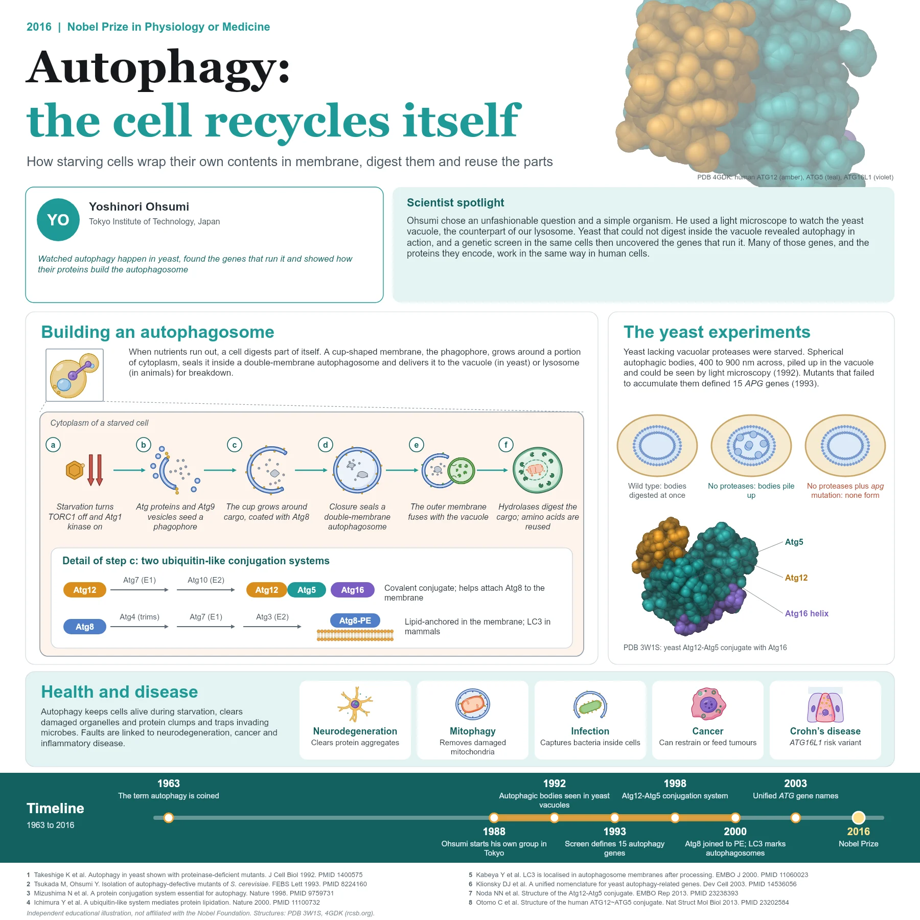
Yoshinori Ohsumi reasoned that the yeast vacuole, the counterpart of the lysosome, might reveal autophagy if its digestion could be stopped. He starved yeast strains that lacked the main vacuolar proteases. Within hours, small spherical bodies, 400 to 900 nanometres across, accumulated in the vacuole and could be seen moving under an ordinary light microscope. Electron microscopy showed that these "autophagic bodies" contained ribosomes, mitochondria and other cytoplasmic material: the vacuole was receiving portions of the cell. The work was published in 1992.
This simple visual read-out made a genetic screen possible. Ohsumi’s group mutagenised the protease-deficient yeast and searched for cells that failed to accumulate autophagic bodies when starved. In 1993 they reported 15 genes needed for autophagy, first called APG genes and later unified as ATG genes. Cloning and characterising these genes over the following years revealed the core machinery, much of which is conserved in human cells.
How the mechanism works
Starvation inactivates the nutrient-sensing kinase complex TORC1 and activates the Atg1 kinase complex (ULK1 in mammals). Atg proteins, small vesicles carrying Atg9 and a phosphatidylinositol 3-kinase complex gather at an assembly site and nucleate a flat, cup-shaped membrane called the phagophore.
The phagophore then expands around its cargo, driven by two ubiquitin-like conjugation systems discovered in Ohsumi’s laboratory. In the first, Atg12 is activated by the E1-like enzyme Atg7, passed to the E2-like Atg10 and joined covalently to Atg5; the Atg12-Atg5 conjugate pairs with Atg16. In the second, Atg8 is trimmed by the protease Atg4, activated by Atg7, handed to Atg3 and linked to the lipid phosphatidylethanolamine, anchoring it in the growing membrane. The Atg12-Atg5 complex helps this lipidation step. The mammalian form of Atg8, LC3, became the standard marker of autophagosomes.
When the edges of the cup meet, the membrane seals into a double-membrane autophagosome. Its outer membrane fuses with the vacuole or lysosome, hydrolases digest the inner membrane and cargo, and amino acids and other building blocks return to the cytoplasm for reuse.
From discovery to applications
Autophagy turned out to be essential far beyond starvation. It removes damaged mitochondria (mitophagy), clears protein aggregates that build up in neurodegenerative diseases, captures bacteria that invade the cytoplasm and helps cells survive stress. Variants in the autophagy gene ATG16L1 are associated with Crohn’s disease, and autophagy can either restrain tumour formation or help established tumours survive, depending on the setting.
Autophagy is not only bulk and non-selective. In selective forms, receptor proteins bind both the cargo, often marked with ubiquitin, and Atg8 family proteins on the phagophore through short LC3-interacting motifs. Damaged mitochondria are removed with the help of PINK1 and Parkin, two proteins linked to inherited Parkinson’s disease, while other receptors target protein clumps or invading bacteria.
The importance of autophagy for the whole body is clear in mice: animals unable to make the Atg12-Atg5 conjugate are born normally but die within a day, during the starvation period that follows birth before suckling begins.
Researchers are now exploring ways to boost or block autophagy with drugs. The conversion of LC3 to its lipid-linked form and the appearance of LC3-labelled dots in cells are standard ways to measure autophagy in cells and tissues.
Why it matters today
Ohsumi’s work shows the power of a simple organism and a clear visual assay. A question that had puzzled cell biologists for decades was opened up by watching yeast vacuoles under a light microscope, and the genes found in that screen now anchor a large field that connects basic cell biology with neurology, immunology and cancer research.
Key takeaways
- Starved protease-deficient yeast accumulated autophagic bodies in the vacuole, making autophagy visible.
- A genetic screen in 1993 identified 15 genes required for autophagy, now called ATG genes.
- Two ubiquitin-like systems, Atg12-Atg5 and Atg8-PE (LC3 in mammals), drive autophagosome formation.
- Autophagy protects against neurodegeneration and infection and is linked to cancer and Crohn’s disease.
References
- Takeshige K, Baba M, Tsuboi S, et al. Autophagy in yeast demonstrated with proteinase-deficient mutants and conditions for its induction. J Cell Biol. 1992;119(2):301-11. PubMed 1400575
- Tsukada M, Ohsumi Y. Isolation and characterization of autophagy-defective mutants of Saccharomyces cerevisiae. FEBS Lett. 1993;333(1-2):169-74. PubMed 8224160
- Mizushima N, Noda T, Yoshimori T, et al. A protein conjugation system essential for autophagy. Nature. 1998;395(6700):395-8. PubMed 9759731
- Ichimura Y, Kirisako T, Takao T, et al. A ubiquitin-like system mediates protein lipidation. Nature. 2000;408(6811):488-92. PubMed 11100732
- Kabeya Y, Mizushima N, Ueno T, et al. LC3, a mammalian homologue of yeast Apg8p, is localized in autophagosome membranes after processing. EMBO J. 2000;19(21):5720-8. PubMed 11060023
- Klionsky DJ, Cregg JM, Dunn WA Jr, et al. A unified nomenclature for yeast autophagy-related genes. Dev Cell. 2003;5(4):539-45. PubMed 14536056
- Noda NN, Fujioka Y, Hanada T, et al. Structure of the Atg12-Atg5 conjugate reveals a platform for stimulating Atg8-PE conjugation. EMBO Rep. 2013;14(2):206-11. PubMed 23238393
- Otomo C, Metlagel Z, Takaesu G, et al. Structure of the human ATG12~ATG5 conjugate required for LC3 lipidation in autophagy. Nat Struct Mol Biol. 2013;20(1):59-66. PubMed 23202584
An independent educational article and illustration by BioGein, not affiliated with or endorsed by the Nobel Foundation. Protein structures are rendered from Protein Data Bank entries (rcsb.org).