The question: how does DNA stay intact?
DNA carries the instructions for every cell, and it is copied billions of times over a human lifetime. For a long time it was assumed to be a very stable molecule. If that were true, a cell would need little maintenance. The work honoured in 2015 showed the opposite: DNA is damaged all the time, and life depends on constant repair.
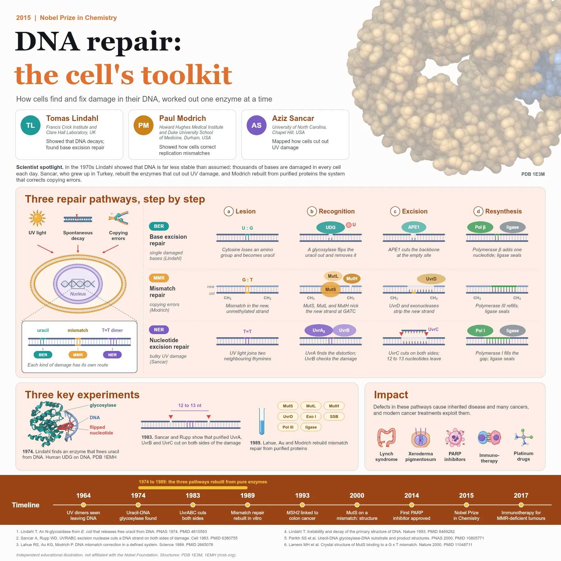
Damage comes from three main sources. Water and oxygen inside the cell attack bases and the sugar backbone, so bases are lost or chemically changed. Ultraviolet light from the sun fuses neighbouring bases. And DNA polymerases, accurate as they are, occasionally insert the wrong base when they copy the genome.
The discovery story
In the early 1970s Tomas Lindahl measured how fast DNA decays and concluded that it is far too unstable to last without repair. One common reaction is the loss of an amino group from cytosine, which turns it into uracil, a base that normally belongs in RNA. In 1974 he found an enzyme in Escherichia coli, uracil-DNA glycosylase, that removes uracil from DNA. This was the first step of a pathway now called base excision repair. In a 1993 review he estimated that thousands of potentially harmful changes occur in the DNA of each human cell every day.
Aziz Sancar, who grew up in Turkey and trained as a doctor before turning to biochemistry, studied how bacteria survive ultraviolet light. In 1983, working with W. Dean Rupp, he showed that three purified proteins, UvrA, UvrB and UvrC, together cut a damaged DNA strand on both sides of a UV lesion. This defined the core of nucleotide excision repair.
Paul Modrich asked how cells correct the mistakes made when DNA is copied. In bacteria, newly made DNA is briefly unmethylated at GATC sequences, which lets the repair system tell the new strand from the old one. In 1989 his group rebuilt mismatch repair in a test tube from purified proteins, proving that a defined set of enzymes is enough to find a mismatch and correct the right strand.
How the three pathways work
Base excision repair handles small changes to single bases. A DNA glycosylase flips the damaged base out of the helix and cuts it from its sugar, leaving an abasic site. The enzyme APE1 cuts the backbone at that site, DNA polymerase beta inserts the correct nucleotide, and DNA ligase seals the strand. Structures such as human uracil-DNA glycosylase bound to DNA (PDB entry 1EMH) show the flipped-out nucleotide inside the enzyme.
Mismatch repair corrects copying errors. In Escherichia coli, the protein MutS recognises a mismatched base pair, as seen in the structure of MutS bound to a G:T mismatch (PDB entry 1E3M). MutL joins it and activates MutH, which nicks the unmethylated new strand at a nearby GATC site. The helicase UvrD and exonucleases then remove the new strand past the error, DNA polymerase III fills the gap and ligase seals it.
Nucleotide excision repair removes bulky lesions such as UV-induced thymine dimers. In bacteria, UvrA finds the distortion in the helix and loads UvrB, which verifies the damage. UvrC then cuts on both sides, and a fragment of 12 to 13 nucleotides is released. DNA polymerase I fills the gap and ligase seals it. Human cells use a related but more complex set of proteins and remove a longer fragment.
From discovery to medicine
Repair defects cause disease. People with xeroderma pigmentosum lack nucleotide excision repair and are extremely sensitive to sunlight. In 1993 the human MSH2 gene, a relative of mutS, was linked to an inherited form of colon cancer now called Lynch syndrome.
Cancer therapy also builds on repair biology. Platinum drugs such as cisplatin create DNA adducts that cells must remove. PARP inhibitors, first approved in 2014, are especially toxic to tumour cells that already lack other repair routes. And in 2017 an immune checkpoint antibody was approved for tumours with defective mismatch repair, which carry many mutations that the immune system can recognise.
Why it matters today
The 2015 prize shows the value of patient biochemistry: isolating one enzyme at a time and rebuilding a process from pure parts. The resulting picture explains why genomes remain stable, why some people are born with a high risk of cancer, and how modern treatments can turn a tumour's repair weakness against it.
Key takeaways
- DNA is chemically unstable, and thousands of changes arise in each human cell every day.
- Base excision repair, started by DNA glycosylases, fixes single damaged bases such as uracil.
- Mismatch repair uses MutS, MutL and MutH to find copying errors and remove the newly made strand.
- Nucleotide excision repair cuts out UV damage on both sides, releasing a short fragment.
- Repair defects cause inherited diseases and cancers, and several cancer treatments exploit them.
References
- Lindahl T. An N-glycosidase from Escherichia coli that releases free uracil from DNA containing deaminated cytosine residues. Proc Natl Acad Sci U S A. 1974;71(9):3649-53. PubMed 4610583
- Sancar A, Rupp WD. A novel repair enzyme: UVRABC excision nuclease of Escherichia coli cuts a DNA strand on both sides of the damaged region. Cell. 1983;33(1):249-60. PubMed 6380755
- Lahue RS, Au KG, Modrich P. DNA mismatch correction in a defined system. Science. 1989;245(4914):160-4. PubMed 2665076
- Lindahl T. Instability and decay of the primary structure of DNA. Nature. 1993;362(6422):709-15. PubMed 8469282
- Fishel R, Lescoe MK, Rao MR, et al. The human mutator gene homolog MSH2 and its association with hereditary nonpolyposis colon cancer. Cell. 1993;75(5):1027-38. PubMed 8252616
- Parikh SS, Walcher G, Jones GD, et al. Uracil-DNA glycosylase-DNA substrate and product structures: conformational strain promotes catalytic efficiency by coupled stereoelectronic effects. Proc Natl Acad Sci U S A. 2000;97(10):5083-8. PubMed 10805771
- Lamers MH, Perrakis A, Enzlin JH, et al. The crystal structure of DNA mismatch repair protein MutS binding to a G x T mismatch. Nature. 2000;407(6805):711-7. PubMed 11048711
An independent educational article and illustration by BioGein, not affiliated with or endorsed by the Nobel Foundation. Protein structures are rendered from Protein Data Bank entries (rcsb.org).