The question: how does cargo reach the right place at the right time?
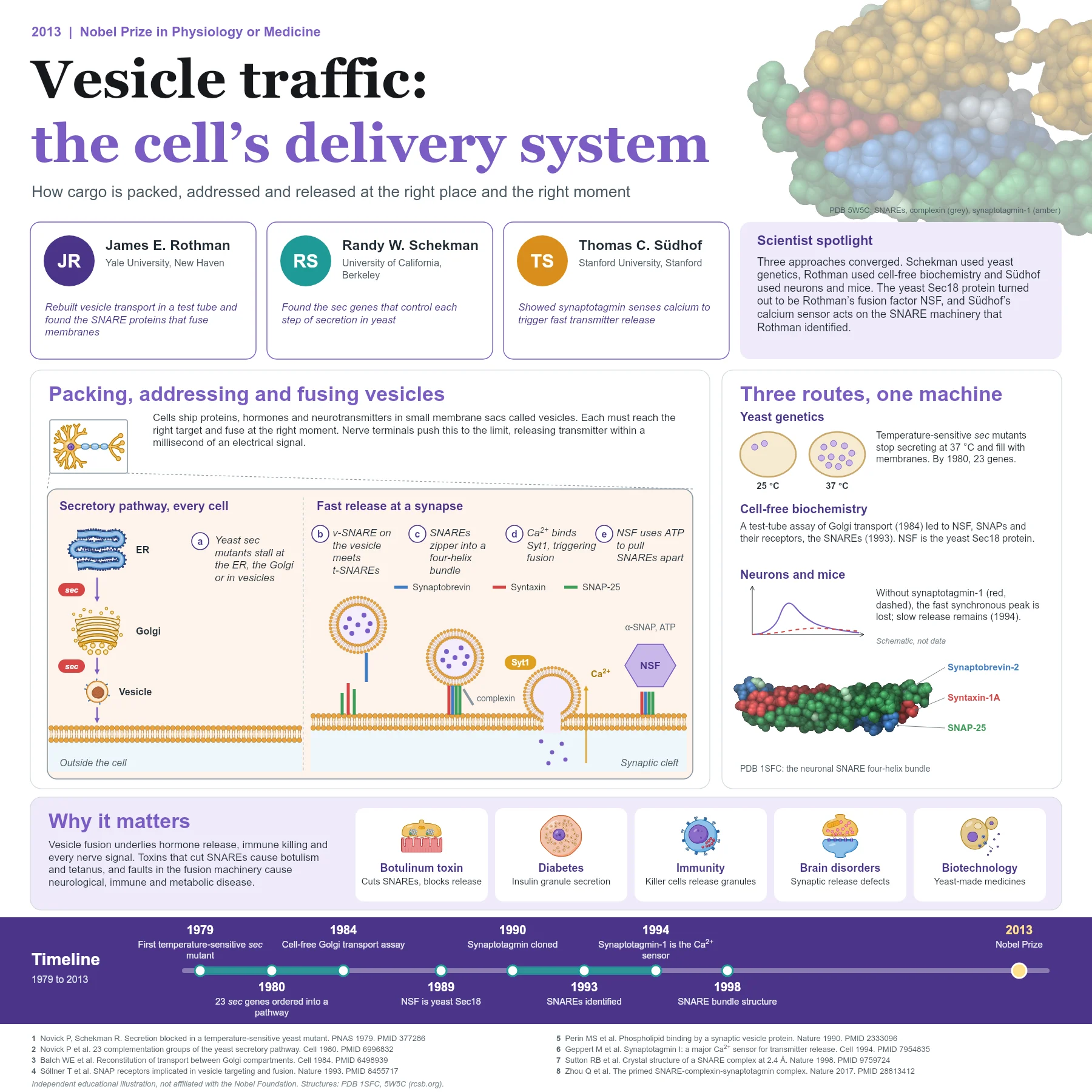
Cells make proteins, hormones and neurotransmitters in one place and need them somewhere else. Much of this cargo travels in vesicles, tiny membrane sacs that bud from one compartment and fuse with another: from the endoplasmic reticulum (ER) to the Golgi apparatus, from the Golgi to the cell surface, and, in nerve terminals, from synaptic vesicles to the synaptic cleft.
Two problems must be solved. Each vesicle has to recognise its correct target among many membranes, and fusion has to happen at the right moment, sometimes within a millisecond of a signal. The 2013 laureates found the genes and proteins that solve both.
Earlier pulse-chase experiments had already traced newly made secretory proteins from the ER through the Golgi to secretory granules. That work defined the route, but not the molecules that drive vesicles along it or decide where they fuse.
The discovery story
Randy Schekman used yeast genetics. In 1979 he and his student reported a temperature-sensitive mutant that stopped secreting at 37 °C and accumulated internal membranes. By 1980 a larger screen had identified 23 groups of sec genes, and the type of membrane that piled up in each mutant revealed where in the pathway the gene acted: the ER, the Golgi or secretory vesicles.
James Rothman took a biochemical route. In 1984 his group reconstituted transport between Golgi compartments in a test tube, which let them purify the proteins needed for fusion. They identified NSF, an ATPase, and its adaptor proteins, the SNAPs, and in 1993 used them as bait to fish out their membrane receptors, the SNAREs. One SNARE sits on the vesicle and others on the target membrane, and their specific pairing suggested how a vesicle docks at the right place. The two approaches met when the yeast gene SEC18 proved to encode NSF.
Thomas Südhof studied how neurons release transmitter so quickly. His group cloned synaptotagmin, a synaptic vesicle protein with calcium and phospholipid binding domains, and in 1994 showed that neurons from mice lacking synaptotagmin-1 lose fast, synchronous release, while slower release remains. Synaptotagmin-1 is the main calcium sensor that couples the electrical signal to vesicle fusion. Südhof’s group also studied other release proteins, including Munc18-1 and complexin, showing how they cooperate with the SNAREs and synaptotagmin.
How the mechanism works
At a nerve terminal, the vesicle SNARE synaptobrevin (VAMP2) meets the plasma membrane SNAREs syntaxin-1 and SNAP-25. Their helices zip together from the membrane-distal end towards the membranes, forming a tight four-helix bundle that pulls the two bilayers into contact. The crystal structure of this core complex (PDB 1SFC) shows four parallel helices about 12 nanometres long.
Partly zipped complexes are held ready by proteins such as complexin and Munc18. When an action potential opens voltage-gated calcium channels, calcium binds synaptotagmin-1, which interacts with membranes and the SNARE complex and triggers fusion pore opening, releasing transmitter into the cleft. Afterwards, NSF and α-SNAP use ATP to pull the spent SNARE complexes apart so the proteins can be reused. A structure of the primed SNARE, complexin and synaptotagmin complex (PDB 5W5C) shows how these parts fit together before release.
Earlier in the pathway, vesicles bud from the ER with the help of coat proteins: Schekman’s group later showed that a set of sec gene products forms the COPII coat that shapes ER-derived vesicles. Different SNARE combinations operate at each step of traffic, which helps keep compartments distinct while membranes and cargo flow between them.
From discovery to applications
The same machinery operates throughout the body. Insulin-secreting beta cells, hormone-producing glands and immune cells that release cytotoxic granules all rely on SNAREs and calcium sensors, and inherited defects in fusion proteins cause immune, neurological and metabolic disorders. Botulinum and tetanus neurotoxins act by cutting SNARE proteins, which explains their potency and underlies the medical use of botulinum toxin to relax overactive muscles. Knowledge of the yeast secretory pathway also supports biotechnology, where yeast is used to make medicines such as insulin.
Why it matters today
This prize illustrates how genetics, biochemistry and physiology can converge on one answer. Yeast mutants, test-tube reactions and mouse neurons each revealed part of the same machine, and together they explain how cells keep their compartments organised and how the brain sends signals with millisecond precision.
Key takeaways
- Yeast sec mutants revealed the genes that control successive steps of secretion.
- A cell-free transport assay led to NSF, SNAPs and the SNARE proteins that fuse membranes.
- Vesicle and target SNAREs zip into a four-helix bundle that drives fusion.
- Synaptotagmin-1 senses calcium to trigger fast, synchronous neurotransmitter release.
References
- Novick P, Schekman R. Secretion and cell-surface growth are blocked in a temperature-sensitive mutant of Saccharomyces cerevisiae. Proc Natl Acad Sci U S A. 1979;76(4):1858-62. PubMed 377286
- Novick P, Field C, Schekman R. Identification of 23 complementation groups required for post-translational events in the yeast secretory pathway. Cell. 1980;21(1):205-15. PubMed 6996832
- Balch WE, Dunphy WG, Braell WA, et al. Reconstitution of the transport of protein between successive compartments of the Golgi measured by the coupled incorporation of N-acetylglucosamine. Cell. 1984;39(2 Pt 1):405-16. PubMed 6498939
- Wilson DW, Wilcox CA, Flynn GC, et al. A fusion protein required for vesicle-mediated transport in both mammalian cells and yeast. Nature. 1989;339(6223):355-9. PubMed 2657434
- Söllner T, Whiteheart SW, Brunner M, et al. SNAP receptors implicated in vesicle targeting and fusion. Nature. 1993;362(6418):318-24. PubMed 8455717
- Perin MS, Fried VA, Mignery GA, et al. Phospholipid binding by a synaptic vesicle protein homologous to the regulatory region of protein kinase C. Nature. 1990;345(6272):260-3. PubMed 2333096
- Geppert M, Goda Y, Hammer RE, et al. Synaptotagmin I: a major Ca2+ sensor for transmitter release at a central synapse. Cell. 1994;79(4):717-27. PubMed 7954835
- Sutton RB, Fasshauer D, Jahn R, et al. Crystal structure of a SNARE complex involved in synaptic exocytosis at 2.4 A resolution. Nature. 1998;395(6700):347-53. PubMed 9759724
- Zhou Q, Zhou P, Wang AL, et al. The primed SNARE-complexin-synaptotagmin complex for neuronal exocytosis. Nature. 2017;548(7668):420-425. PubMed 28813412
An independent educational article and illustration by BioGein, not affiliated with or endorsed by the Nobel Foundation. Protein structures are rendered from Protein Data Bank entries (rcsb.org).