The question: is specialisation permanent?
A fertilised egg gives rise to hundreds of cell types, from neurons to muscle. For much of the twentieth century it was debated whether specialised cells lose the genes they no longer use, or keep them in a silent but recoverable form. If the full genome is retained, it might be possible to reverse development and turn a mature cell back into one that can form any tissue.
Development was often pictured as a ball rolling downhill into ever narrower valleys, each valley a cell fate. In that picture specialisation looks like a one-way journey. The two laureates showed that the ball can be pushed back to the top.
The discovery story
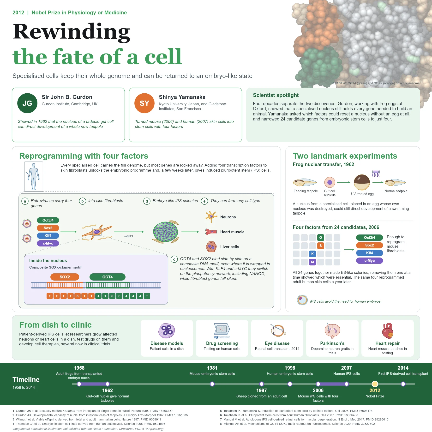
John Gurdon tested this with frogs. Working at Oxford with eggs of Xenopus laevis, he destroyed the egg’s own nucleus with ultraviolet light and replaced it with a nucleus taken from another cell. In 1958 he reported fertile adult frogs from nuclei of embryonic cells. In 1962 he went further and used nuclei from intestinal cells of feeding tadpoles, which are specialised; some of these eggs developed into normal swimming tadpoles. A specialised nucleus therefore still contained all the information needed to build an animal. Later cloning of mammals, such as the sheep produced from an adult cell in 1997, built on the same principle.
Shinya Yamanaka asked whether the egg could be replaced by a few defined factors. Using knowledge of genes active in embryonic stem cells, his group selected 24 candidate transcription factors and introduced them together into mouse fibroblasts with retroviruses, selecting for cells that activated an embryonic stem cell reporter. Colonies resembling embryonic stem cells appeared. By removing one factor at a time they found that four were enough: Oct3/4, Sox2, Klf4 and c-Myc. The resulting induced pluripotent stem (iPS) cells were reported in 2006, and in 2007 the same four factors reprogrammed adult human skin cells.
How reprogramming works
The four factors are transcription factors. OCT4 and SOX2 bind side by side on composite DNA motifs, combining a SOX element with the octamer sequence ATGCAAAT. They can engage their targets even when the DNA is wrapped around nucleosomes, as shown in the structure of OCT4 and SOX2 bound to a nucleosome (PDB 6T90), which helps them open silent chromatin. Together with KLF4 and c-MYC they activate the pluripotency network, including the cell’s own OCT4 and NANOG genes, while the fibroblast programme is gradually shut down.
Reprogramming is slow and inefficient: over a few weeks only a small fraction of cells complete the transition, changing shape, switching metabolism and resetting their epigenetic marks. The successful cells form compact colonies that can grow indefinitely and differentiate into cell types of all three germ layers, including neurons, heart muscle and liver cells.
From discovery to applications
iPS cells can be made from any person, which means patient-specific cells can be grown in the laboratory. Researchers use them to model diseases, for example neurons from people with inherited neurological disorders, and to test drugs on human cells. Because they do not require human embryos, iPS cells also avoided an ethical barrier faced by embryonic stem cell research.
Clinical use began in 2014, when retinal pigment epithelium cells derived from a patient’s own iPS cells were transplanted to treat macular degeneration. Trials of iPS cell-derived dopamine neurons for Parkinson’s disease and heart muscle for heart failure are under way, with careful attention to safety.
Safety drove further refinements. c-Myc is an oncogene and retroviruses insert permanently into the genome, so later methods omitted c-Myc or delivered the factors without integration, for example with Sendai virus, episomal plasmids or synthetic mRNA. In 2007 an independent group also reported human iPS cells made with a different set of factors, OCT4, SOX2, NANOG and LIN28, confirming that reprogramming is robust.
Why it matters today
Together the two discoveries changed how biologists think about cell identity: it is maintained actively and can be reset. Gurdon’s nuclear transfer experiments established the principle, and Yamanaka’s four factors turned it into a practical tool that is now used in laboratories worldwide.
Key takeaways
- Nuclear transfer in frogs showed that specialised cells keep a complete genome.
- Four transcription factors, Oct3/4, Sox2, Klf4 and c-Myc, reprogram fibroblasts into iPS cells.
- OCT4 and SOX2 bind composite DNA motifs, even on nucleosomes, to switch on the pluripotency network.
- iPS cells support disease modelling, drug testing and early cell therapies.
References
- Gurdon JB, ELSDALE TR, FISCHBERG M. Sexually mature individuals of Xenopus laevis from the transplantation of single somatic nuclei. Nature. 1958;182(4627):64-5. PubMed 13566187
- Gurdon JB. The developmental capacity of nuclei taken from intestinal epithelium cells of feeding tadpoles. J Embryol Exp Morphol. 1962;10:622-40. PubMed 13951335
- Wilmut I, Schnieke AE, McWhir J, et al. Viable offspring derived from fetal and adult mammalian cells. Nature. 1997;385(6619):810-3. PubMed 9039911
- Thomson JA, Itskovitz-Eldor J, Shapiro SS, et al. Embryonic stem cell lines derived from human blastocysts. Science. 1998;282(5391):1145-7. PubMed 9804556
- Takahashi K, Yamanaka S. Induction of pluripotent stem cells from mouse embryonic and adult fibroblast cultures by defined factors. Cell. 2006;126(4):663-76. PubMed 16904174
- Takahashi K, Tanabe K, Ohnuki M, et al. Induction of pluripotent stem cells from adult human fibroblasts by defined factors. Cell. 2007;131(5):861-72. PubMed 18035408
- Mandai M, Watanabe A, Kurimoto Y, et al. Autologous Induced Stem-Cell-Derived Retinal Cells for Macular Degeneration. N Engl J Med. 2017;376(11):1038-1046. PubMed 28296613
- Michael AK, Grand RS, Isbel L, et al. Mechanisms of OCT4-SOX2 motif readout on nucleosomes. Science. 2020;368(6498):1460-1465. PubMed 32327602
An independent educational article and illustration by BioGein, not affiliated with or endorsed by the Nobel Foundation. Protein structures are rendered from Protein Data Bank entries (rcsb.org).