The question: how does a hormone outside a cell change what happens inside?
Adrenaline released into the blood makes the heart beat faster and opens the airways, yet the hormone itself does not enter the cells it acts on. In the middle of the twentieth century the idea of a receptor, a molecule on the cell surface that recognises the hormone, was widely used but the receptors themselves had never been isolated.
The 2012 Nobel Prize in Chemistry went to Robert Lefkowitz and Brian Kobilka for studies of G-protein-coupled receptors (GPCRs), the largest family of receptors in the human body. They showed what these receptors are, how they are built and how they pass a signal through the membrane.
The discovery story
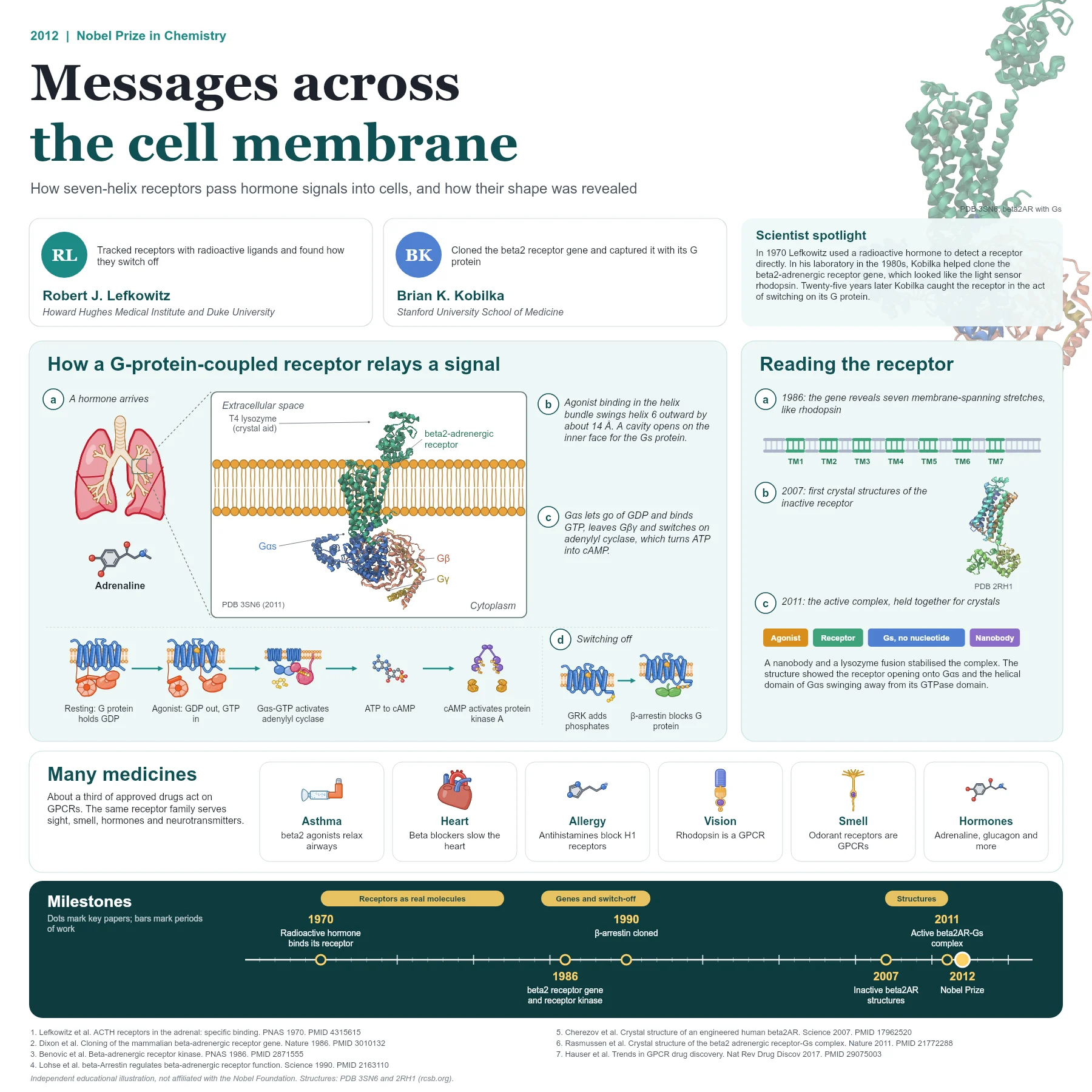
In 1970 Lefkowitz and colleagues attached radioactive iodine to the hormone ACTH and showed that it bound specifically to adrenal tissue in proportion to biological activity. Radioactive ligands became his way to find and count receptors, and his laboratory went on to purify the beta-adrenergic receptors that respond to adrenaline.
In 1986 the team, with Kobilka as a key member, cloned the gene and cDNA of the mammalian beta2-adrenergic receptor. Its sequence resembled rhodopsin, the light receptor of the eye, and suggested several stretches that cross the membrane. This similarity revealed a family: receptors for hormones, neurotransmitters and light share a seven-helix design.
The same year Lefkowitz's group identified the beta-adrenergic receptor kinase, which phosphorylates only the agonist-occupied receptor, and in 1990 they cloned beta-arrestin, a protein that binds the phosphorylated receptor and stops it from signalling. This explained how cells become desensitised after prolonged stimulation.
Beta-arrestin turned out to be a 418-amino-acid protein related to the arrestin of the retina. In test-tube experiments it inhibited signalling by receptors that had been phosphorylated by the receptor kinase by more than 75 percent, which made the switch-off mechanism concrete.
How the mechanism works
A GPCR is a single protein chain that crosses the membrane seven times. An agonist such as adrenaline binds in a pocket among the helices on the outer side. This shifts the helices so that a cavity opens on the inner face of the receptor.
A heterotrimeric G protein, made of alpha, beta and gamma subunits, docks into that cavity. The receptor makes the alpha subunit release GDP and take up GTP. For the beta2-adrenergic receptor the G protein is Gs: the GTP-bound alpha subunit separates from the beta-gamma pair and activates adenylyl cyclase, which converts ATP into the second messenger cyclic AMP. Cyclic AMP then activates protein kinase A, which changes the activity of many proteins in the cell.
To switch the signal off, G-protein-coupled receptor kinases add phosphates to the active receptor and beta-arrestin binds, blocking further coupling to the G protein.
Seeing the receptor at work
Receptors are scarce, flexible and unstable outside the membrane, which made them very hard to crystallise. Kobilka spent years engineering the beta2-adrenergic receptor. In 2007 his team and collaborators reported its first crystal structures, one with an antibody fragment and one in which T4 lysozyme replaced a flexible loop (PDB 2RH1).
In 2011 they captured the active complex: an agonist-bound receptor holding a nucleotide-free Gs heterotrimer, stabilised by a nanobody and crystallised (PDB 3SN6). The structure showed the cytoplasmic end of transmembrane helix 6 moved outward by 14 Å, and the helical domain of the Galpha-s subunit swung far away from its GTPase domain. It was the first high-resolution view of transmembrane signalling by a GPCR.
The high-resolution 2007 structure, at 2.4 Å, showed the receptor bound to carazolol, a partial inverse agonist, and revealed that cholesterol helped receptors pack together in the crystal. Comparing it with rhodopsin showed that, although ligands sit in a similar place, each receptor has its own binding site, so rhodopsin alone could not serve as a template for the whole family.
Why it matters today
GPCRs are the most studied drug targets. An analysis published in 2017 counted 475 drugs, about 34% of those approved by the US Food and Drug Administration, that act at GPCRs. They include beta2 agonists for asthma, beta blockers for the heart, antihistamines and opioid painkillers.
The structural work begun with the beta2-adrenergic receptor now guides drug design for many other receptors, and the same family underlies vision, smell and taste.
The same analysis counted about 321 agents in clinical trials, around a fifth of them aimed at GPCRs that have no approved drug yet, so the family remains a major source of new medicines.
Key takeaways
- GPCRs are seven-helix membrane receptors that relay signals from hormones, neurotransmitters, light and odours.
- Agonist binding opens a cavity on the inner face where a G protein exchanges GDP for GTP.
- Gs activates adenylyl cyclase to make cAMP; GRK phosphorylation and beta-arrestin switch the receptor off.
- Cloning of the beta2-adrenergic receptor in 1986 revealed its kinship with rhodopsin.
- The 2011 structure of the beta2AR-Gs complex showed signalling across the membrane in atomic detail.
References
- Lefkowitz RJ, Roth J, Pricer W, Pastan I. ACTH receptors in the adrenal: specific binding of ACTH-125I and its relation to adenyl cyclase. Proc Natl Acad Sci U S A. 1970;65(3):745-52. PubMed 4315615
- Dixon RA, Kobilka BK, Strader DJ, et al. Cloning of the gene and cDNA for mammalian beta-adrenergic receptor and homology with rhodopsin. Nature. 1986;321(6065):75-9. PubMed 3010132
- Benovic JL, Strasser RH, Caron MG, Lefkowitz RJ. Beta-adrenergic receptor kinase: identification of a novel protein kinase that phosphorylates the agonist-occupied form of the receptor. Proc Natl Acad Sci U S A. 1986;83(9):2797-801. PubMed 2871555
- Lohse MJ, Benovic JL, Codina J, Caron MG, Lefkowitz RJ. beta-Arrestin: a protein that regulates beta-adrenergic receptor function. Science. 1990;248(4962):1547-50. PubMed 2163110
- Rasmussen SG, Choi HJ, Rosenbaum DM, et al. Crystal structure of the human beta2 adrenergic G-protein-coupled receptor. Nature. 2007;450(7168):383-7. PubMed 17952055
- Cherezov V, Rosenbaum DM, Hanson MA, et al. High-resolution crystal structure of an engineered human beta2-adrenergic G protein-coupled receptor. Science. 2007;318(5854):1258-65. PubMed 17962520
- Rasmussen SG, DeVree BT, Zou Y, et al. Crystal structure of the beta2 adrenergic receptor-Gs protein complex. Nature. 2011;477(7366):549-55. PubMed 21772288
- Hauser AS, Attwood MM, Rask-Andersen M, Schiöth HB, Gloriam DE. Trends in GPCR drug discovery: new agents, targets and indications. Nat Rev Drug Discov. 2017;16(12):829-842. PubMed 29075003
An independent educational article and illustration by BioGein, not affiliated with or endorsed by the Nobel Foundation. Protein structures are rendered from Protein Data Bank entries (rcsb.org).